OPINION

Enabling technologies for the preparation of multifunctional “bullets” for nanomedicine

Martina K, Serpe L, Cavalli R, Cravotto G
About authors

Department of Drug Science & Technology,
Centre for Nanostructured Interfaces and Surfaces (NIS), University of Turin, Turin, Italy

Correspondence should be addressed to: Giancarlo Cravotto
Via P. Giuria 9, 10125 Turin, Italy; ti.otinu@ottovarc.olracnaig

About paper

Funding: The University of Turin is warmly acknowledged for their financial support (Ricerca Locale 2017).
Received: 2018-06-26 Accepted: 2018-08-30 Published online: 2018-12-30
|
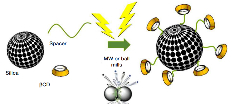
Fig. 1. Schematic representation of cyclodextrin grafted silica prepared under unconventional conditions [6]. Reprinted (adapted) with permission from (Martina K, Baricco F, Berlier G, Caporaso M, Cravotto G. ACS Sustainable Chem. Eng. 2014;2(11):2595-603). Copyright (2018) American Chemical Society)
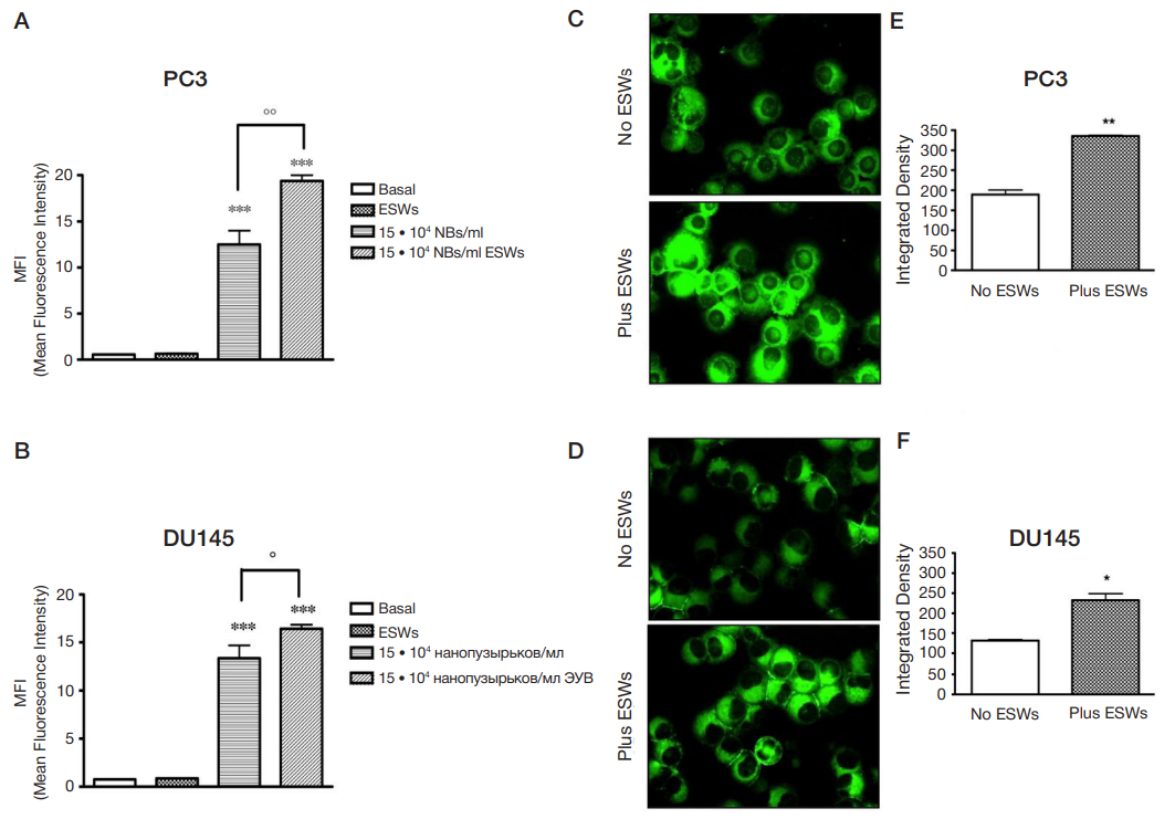
Fig. 2. Nanobubble entrance. Cytofluorimetric analysis of PC3 (A) and DU145 (B) cells treated for 24 hours with 6-coumarin-labelled glycol chitosan NBs (15 • 104 NBs/ml), either in the absence and in the presence of ESWs, expressed as Mean Fluorescence Intensity (MFI). Significance vs untreated cells (Basal): p < 0.001 (* * *); significance vs ESWs: p < 0.05 (°); p < 0.01 (°°). Photos by fluorescence microscope of PC3 (C) and DU145 (D) cells treated with 6-coumarin-labelled glycol chitosan NBs at 15 • 104 NBs/ml, either in the absence and in the presence of ESWs. Pictures were taken at ×200 final magnification (scale bar: 100 μm). The images are representative of three independent experiments; for each experiment, 10 fields were quantified. Image-based quantification of 6-coumarin-labelled glycol chitosan NBs in PC3 (E) and DU145 (F) cells. Significance vs no ESWs: p < 0.05 (*); p < 0.01 (* *). [ref 48]
Table 1. Examples of bioactive molecules loaded into NB formulations