CLINICAL CASE

The new method of pelvic packing against continuing intrapelvic bleeding resulting from the unstable pelvic ring fractures

Egiazaryan KA1, Gordienko DI1, Starchik DA2, Lysko AM1
About authors

1 Pirogov Russian National Research Medical University, Moscow, Russia

2 Mechnikov North-Western State Medical University, Saint Petersburg, Russia

Correspondence should be addressed: Artyom M. Lysko
Khachaturiana 12–3, Moscow, 127562; moc.liamg@oksyLtra

About paper

Author contribution: Egiazaryan KA, Gordienko DI — study organization and planning; Starchik DA — study planning, anatomical examination; Lysko AM — literature analysis, data collection, analysis, interpretation, surgery.
Received: 2019-03-03 Accepted: 2019-04-16 Published online: 2019-04-29
|
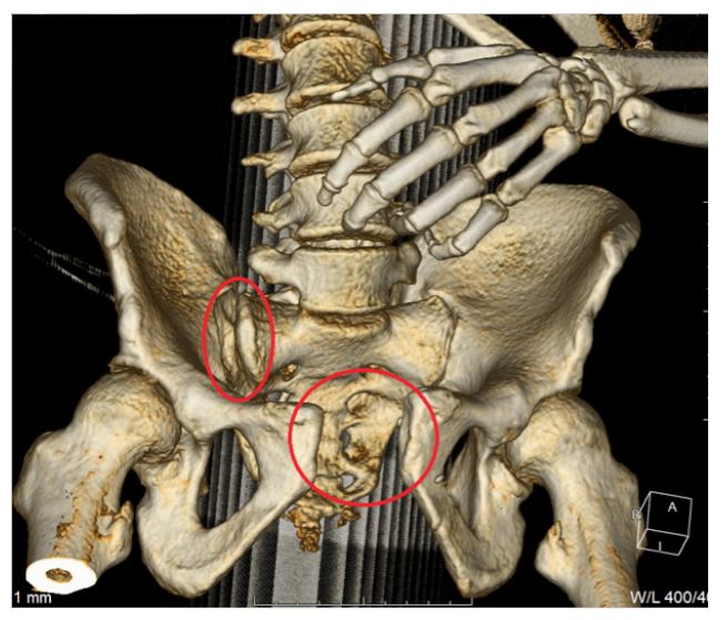
Fig. 1. 3D CT model of the pelvic ring injury: red ovals show the right sacroiliac joint rupture and the pubic symphysis rupture
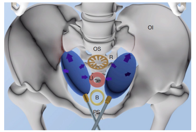
Fig. 2. Schematic representation of the direction of installation of balloon devices in the pelvic cavity: B — bladder, U — uterus, R — rectum, OS — sacrum, Oi — os innominatum, PS — pubic symphysis. The balloons deployed in the lateral space of the pelvis is in blue
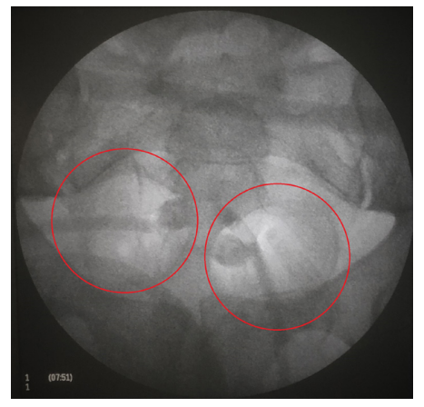
Fig. 3. Intraoperative patient's pelvis X-ray scan, unstable pelvic ring damage and development of intrapelvic hemorrhage after stabilization by external fixation devices and deployment of the balloon devices in the pelvic cavity (balloons circled by red)
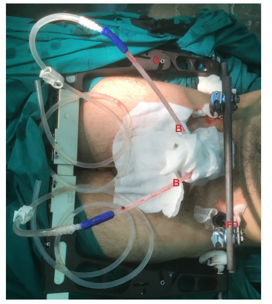
Fig. 4. External appearance of the patient's pelvic region with unstable pelvic ring injury and development of intrapelvic hemorrhage after stabilization by external fixation devices and deployment of the balloon devices in pelvic cavity: С — C-shaped frame for the back of the ring, IFD — External Fixation Device for the front, B — balloon devices
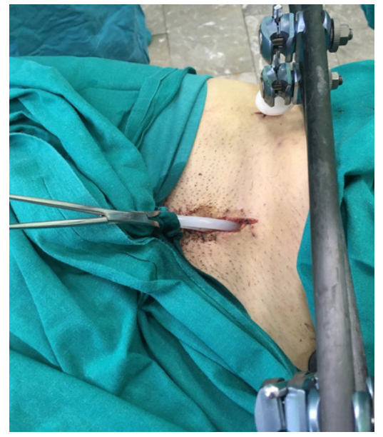
Fig. 5. Appearance of the patient's wound with tubes of balloon devices, before removal, 3rd day. The wound has no signs of inflammation; there is no pathological discharge along the balloon devices. in the right half of the picture — external fixation device on the front of the pelvic ring
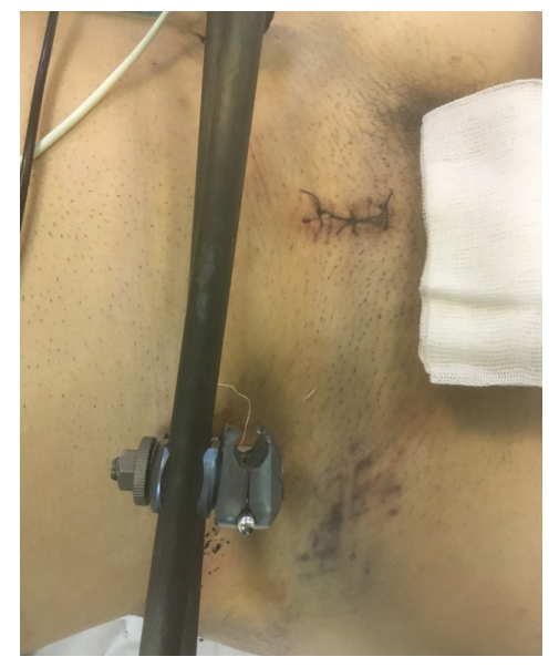
Fig. 6. Postoperative wound, first day after removal of the balloon devices. The postoperative wound heals by primary intention, without signs of inflammation and pathologic discharge; the sutures are consistent. In the left half of the picture — external fixation device on the front of the pelvic ring
Fig. 7. X-rau scan of the patient's pelvis after the final stabilization of the pelvic ring (10th day): right sacroiliac joint fixed with a spongy screw with partial cutting and a washer, pubic symphysis — with plate and screws
Table. Dynamics of the patient's condition indicators before and after surgery