METHOD

A bionic eye: performance of the Argus II retinal prosthesis in low-vision and social rehabilitation of patients with end-stage retinitis pigmentosa

About authors

1 Pirogov Russian National Research Medical University, Moscow, Russia

2 Scientific Clinical Center of Otorhinolaryngology, FMBA of Russia, Moscow, Russia

Correspondence should be addressed: Pavel V. Gliznitsa
Ostrovintyanova 1, Moscow, 117513; moc.duolci@pastinzilg

About paper

Author contribution: Takhchidi KhP — study design, literature analysis, implantation surgery, data analysis and interpretation; Kachclina GF — study design, literature analysis, data analysis and interpretation; Takhchidi NKh — manuscript preparation, literature analysis; Manoyan RA — follow-up observation, rehabilitation sessions, data collection; Gliznitsa PV — follow-up observation, rehabilitation sessions, data collection, manuscript preparation.

Received: 2019-06-04 Accepted: 2019-06-19 Published online: 2019-06-26
|
Fig. 1. OCT scans of patient U.’s right eye before (A) and after (B) implantation of Argus II show the correct position of the implant secured to the retina
Fig. 2. OCT scans of patient Z.’s left eye before (A) and after (B) implantation of Argus II show the correct position of the implant secured to the retina
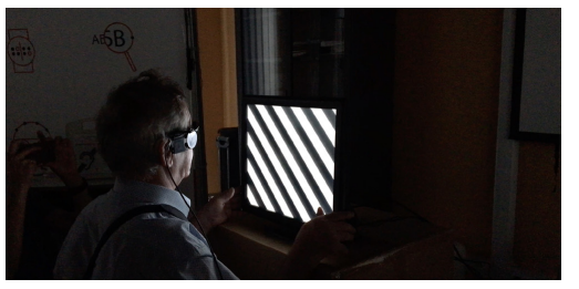
Fig. 3. Patient U. is taking a Grating Visual Acuity test
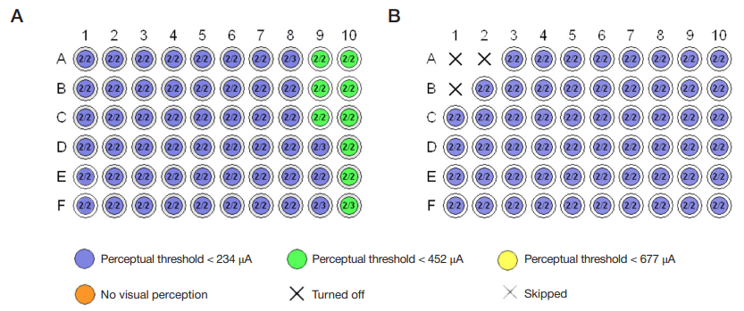
Fig. 4. Stimulation charts of the implants showing perceptual thresholds for each of 60 electrodes in patient U. (A) and patient Z. (B)
Fig. 5. Patient U. identifies a mirror before him and recognizes his own reflection (A). Patient Z. can correctly tell the number of people standing in front of her during the custom fitting session (B)
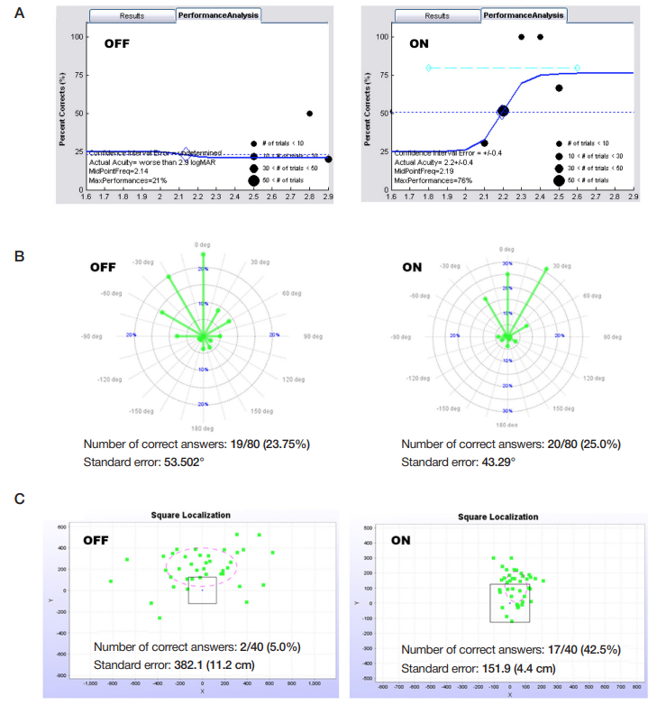
Fig. 6. Results of visual assessment computer tests done by patient U. with Argus II off and on: the Grating Visual Acuity Test (A); the Direction of Motion test (B); the Square Localization test (C)
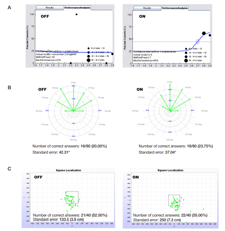
Fig. 7. Results of visual assessment computer tests done by patient Z. with Argus II off and on: the Grating Visual Acuity Test (A); the Direction of Motion test (B); the Square Localization test (C)
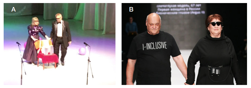
Fig. 8. Patient U. is giving a magic show in Chelyabinsk (A); patient Z. on a catwalk during a fashion show in Moscow (B)