ORIGINAL RESEARCH

Correlated dynamics of serum IGE and IGE+ clonotype count with allergen air level in seasonal allergic rhinitis

About authors

1 Skoltech, Moscow, Russia

2 Shemyakin and Ovchinnikov Institute of Bioorganic Chemistry, Moscow, Russia

3 Pirogov Russian National Research Medical University, Moscow, Russia

Correspondence should be addressed: Ivan V. Zvyagin
Miklukho-Maklaya, 16/10, Moscow, 117997; moc.liamg@nigayvzi

About paper

Funding: the study was supported by the Grants Council under the President of the Russian Federation (grant MK6000.2018.4).

Acknowledgments: we are very grateful to all the donors who participated in the study.

Author contribution: Mikelov AI — antibody levels determination, IGH cDNA libraries preparation, sequencing data and results analysis, research design, drafting; Staroverov DB — cell subpopulations isolation (flow cytofluorometry); Komech EA — blood samples collection, cell subpopulations isolation (flow cytofluorometry); Lebedev YB — results analysis and discussion, advisory support; Chudakov DM — results analysis and discussion, advisory support (cDNA libraries preparation); Zvyagin IV — IGH cDNA libraries preparation,sequencing data and results analysis, research design, drafting, research organization.
Received: 2019-10-09 Accepted: 2019-10-23 Published online: 2019-10-31
|
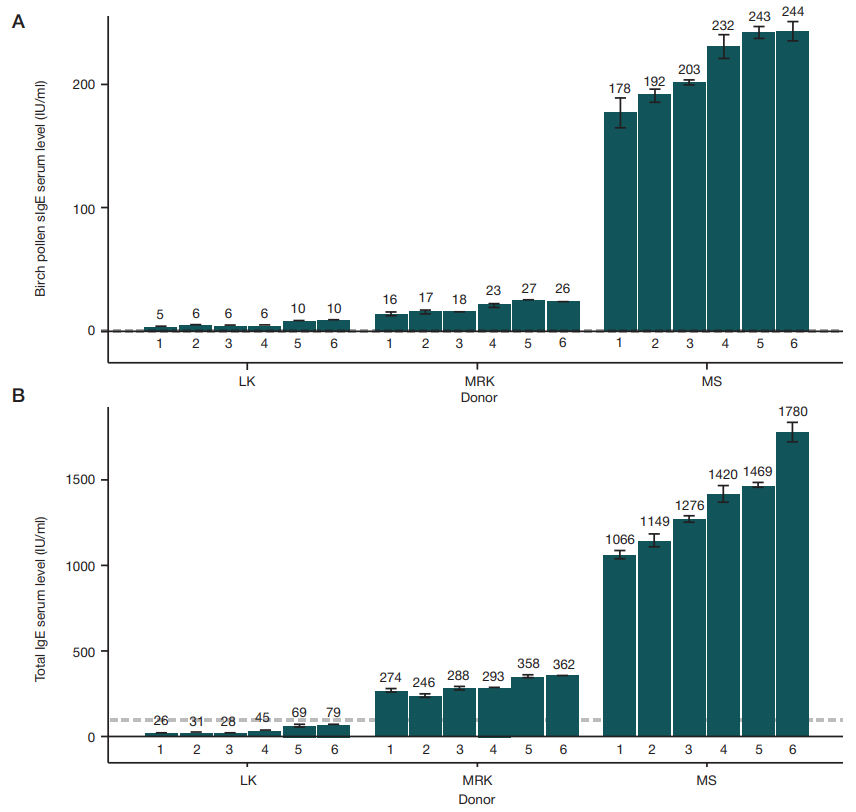
Fig. 1. Level of birch pollen-specific IgE (A) and total IgE (B) for three donors (LK, MRK, MS) with seasonal allergic rhinoconjunctivitis at six time points (1–6). Horizontal dashed line represents the reference level of corresponding antibodies in a healthy adult donor's serum (<0.35 IU/ml for sIgE, <100 IU/ml for general IgE). Error bars reflect difference between minimum and maximum measurement values
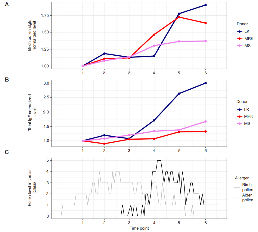
Fig. 2. Serum IgE level and birch pollen air level dynamics. A. Birch pollen sIgE serum level at each time point, normalized to the level of birch pollen sIgE at time point 1 for the corresponding donor. B. Total sIgE serum level at each time point, normalized to the level of total sIgE at time point 1 for the corresponding donor. C. Birch pollen level in the air
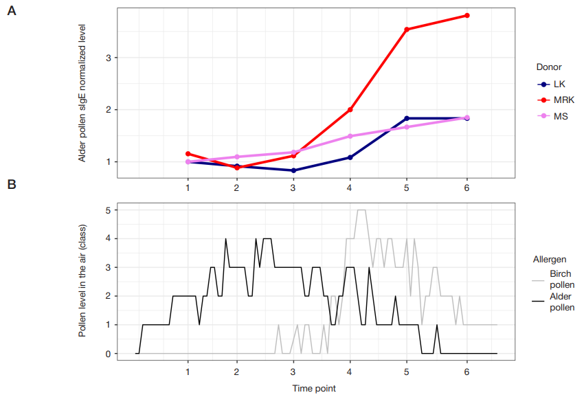
Fig. 3. Dynamics of alder pollen-specific IgE level and concentration of pollen in the air. A. Alder pollen sIgE serum level at each time point, normalized to the level of alder pollen sIgE at time point 1 for the corresponding donor. B. Pollen level in the air
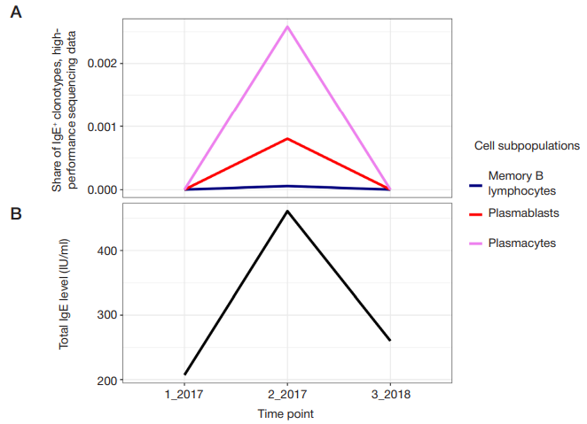
Fig. 4. B cell clonal repertoire dynamics. A. Abundance of IgE-clonotypes as revealed by high-throughput sequencing of immunoglobulin heavy chain repertoires for three subpopulations of peripheral blood cells: memory B lymphocytes, plasmablasts and plasma cells. B. Serum level of total IgE at the corresponding time points
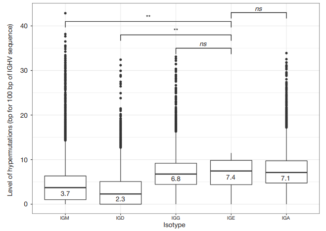
Fig. 5. Level of hypermutations (per 100 bp of IGHV sequence) calculated from high-throughput IGH repertoire sequencing data. IGM (n = 55 865), IGD (n = 15 834), IGG (n = 20 321), IGE (n = 13), IGA (n = 23 955). (* * — p < 0.01; ns — not significant; Wilcoxon rank-sum test)
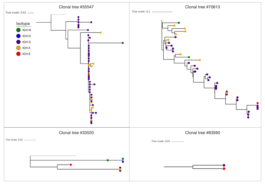
Fig. 6. Phylogenetic trees for IgE-containing clonal groups with clonotypes of different isotypes. Each dot represents a unique IGH clonotype. Clonotypes of different isotypes are color coded (dark blue — IgG, red — IgE, orange — IgA, green — IgM, blue — IgD). Length of the branches reflects distance between nodes according to the number of mutations between sequences. "Tree scale" reflects the scale used to build the tree
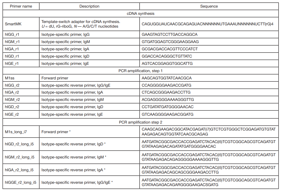
Table 1. Sequences of oligonucleotide primers used for IGH cDNA libraries
Note: * (i5) and (i7) — sequences 8 nucleotides in length, which are sample-specific barcodes.
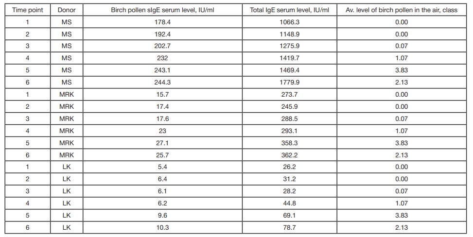
Table 2. Data used to build the regression models. Birch pollen-specific and total IgE levels, averaged level of birch pollen in the air between corresponding time points
Table 3. Pollination level — pollination classes according to the volume of pollen in the air