ORIGINAL RESEARCH

The impact of the novel coronavirus infection COVID-19 on the mother-placenta-fetus system

About authors

Kulakov National Medical Research Center for Obstetrics, Gynecology and Perinatology, Moscow, Russia

Correspondence should be addressed: Natalia A. Lomova
Oparina, 4, Moscow, 117997; ur.xednay@avomol-ahsatan

About paper

Funding: the study was supported by RFBR grant № 20-04-60093.

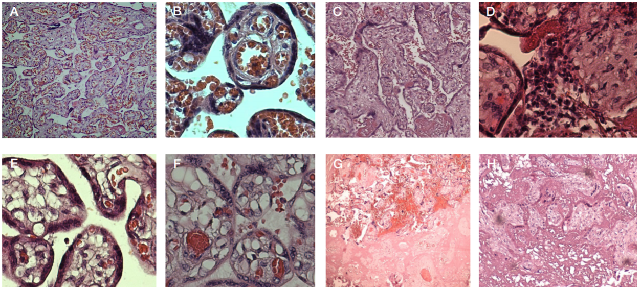
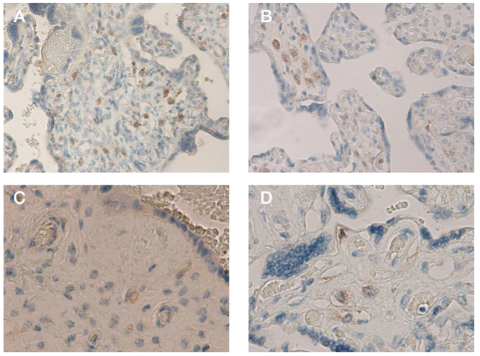
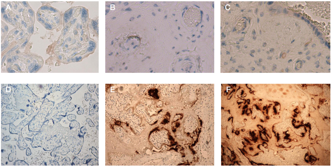
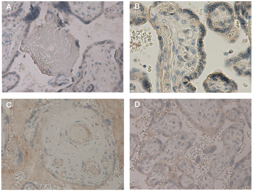
Acknowlegements: here we would like to thank Sinitsina VA, medical laboratory assistant of the 2nd Anatomical Pathology Department of the Kulakov National Medical Research Center for Obstetrics, Gynecology and Perinatology, and Bugrova AV, senior research scientist of the Laboratory for Proteomics and Metabolomics of Human Reproduction of the Kulakov National Medical Research Center for Obstetrics, Gynecology and Perinatology.

Author contribution: Nizyaeva NV — pathomorphological study and IHC analysis, systematic analysis, manuscript writing; Lomova NA — analysis of clinical data, systematic analysis, manuscript writing; Dolgopolova EL — collection and preparation of biological matrix samples in the red zone, statistical analysis of the results; Petrova UL — collection and preparation of biological matrix samples in the red zone; Karapetyan ТE — analysis of clinical data; Shmakov RG — analysis of clinical data in the red zone, systematic analysis, manuscript editing; Frankevich VE — preparation of the study, systematic analysis.

Compliance with ethical standards: all patients submitted the informed consent to participate in the study; the study met the requirements of the Declaration of Helsinki, International Conference on Harmonization (ICF), Good Clinical Practice (GCP), and Federal Law No. 323-FZ “On the Basics of Protecting Citizens' Health in the Russian Federation” of November 21, 2011.

Received: 2021-04-14 Accepted: 2021-04-25 Published online: 2021-04-30
|
Fig. 1. Histological changes in placental villous trees of women with novel coronavirus infection. А. Placenta of women with uncomplicated pregnancies, one terminal villus contains 3–7 vessels; ×40. B. Placenta of women with uncomplicated pregnancies, one terminal villus contains 3–7 vessels 3–7; ×400. C. Placenta of patients with Covid-19; increased fibrinoid deposition; ×100. D. Placenta of patients with Covid-19; focal accumulations of inflammatory cells (macrophages, lymphocytes with a dash of plasmocytes and sporadic neutrophils); ×400. E. Placenta of patients with Covid-19; angiogenesis with predominant vascular branching: capillary number increase in one villus to more than 10 together with blood vessel diameter reduction; intact syncytiotrophoblast membranes attract attention. F. Placenta of patients with Covid-19; angiogenesis with predominant vascular branching: capillary number increase in one villus to more than 10 together with blood vessel diameter reduction. G. Placenta of patients with Covid-19; multiple infarction and hemorrhages in placental samples obtained from women with Covid-19. H. Placenta of patients with Covid-19
Fig. 2. CD4 expression in placental tissue in patients with Covid-19 and comparison group. А. Focal increase in the number of СD4+ macrophages and lymphocytes in the villous stroma in patients with Сovid-19; ×200. B. Focal increase in the number of СD4+ macrophages and lymphocytes in the villous stroma in patients with Сovid-19; ×200. C. Focal increase in the number of СD4+ macrophages and lymphocytes in the villous stroma in patients with Сovid-19; ×400. D. Macrophages СD4+ in the villous stroma in women with uncomplicated pregnancy; ×400
Fig. 3. Placental tissue immunohistochemical analysis using primary antibody to SARS-COV-2. А. Weak syncytiotrophoblast membrane and fibrinoid deposits staining in placental tissue of patients with Covid-19, intact syncytiotrophoblast membrane attracts attention, ×200. B. Weak focal staining of endothelium in individual villous vessels; ×400. C. Weak syncytiotrophoblast membrane and fibrinoid deposits staining in placental tissue of patients with Covid-19, intact syncytiotrophoblast membrane attracts attention, ×200; weak focal staining of endothelium in individual villous vessels; ×400. D. Negative staining. Placenta of women with no history of Covid-19; ×100. E. Prominent positive staining of viral particle accumulations around the perimeter of dystrophic villus. Placenta of a woman with acute leukemia, who died from Covid-19. The fetus died in utero from placental insufficiency at 20 weeks of gestation. Stained inflammatory cells are visible: lymphocytes, macrophages; ×100. F. Prominent positive staining of viral particle accumulations around the perimeter of dystrophic villus. Placenta of a woman with acute leukemia, who died from Covid-19. The fetus died in utero at 20 weeks of gestation from placental insufficiency; ×40
Fig. 4. TNFα expression in placental tissue in patients with Covid-19 and comparison group. А. TNFα expression in placental structures of women with uncomplicated pregnancy, weak staining of syncytiotrophoblast membrane and cytoplasm, as well as of vascular endothelium; no staining of fibrinoid deposits; ×100. B. TNFα expression in placental structures of women with uncomplicated pregnancy, weak staining of syncytiotrophoblast membrane and cytoplasm, as well as of vascular endothelium; no staining of fibrinoid deposits; ×200. C. TNFα expression in placental structures of women with Covid-19. Weak staining of vascular endothelium; positively stained fibrinoid deposits; ×200. D. TNFα expression in placental structures of women with Covid-19. Weak staining of vascular endothelium (Fig. C); as well as of syncytiotrophoblast membrane and cytoplasm, and syncytial knots (Fig. D); positively stained fibrinoid deposits (Fig. C); ×200
Fig. 5. IL8 expression in placental tissue in patients with Covid-19 and comparison group. А. IL8 expression in placental structures of women with uncomplicated pregnancy, weak staining of syncytiotrophoblast membrane and cytoplasm, as well as of vascular endothelium; no staining of fibrinoid deposits; ×100. B. IL8 expression in placental structures of women with uncomplicated pregnancy, weak staining of syncytiotrophoblast membrane and cytoplasm, as well as of vascular endothelium; no staining of fibrinoid deposits; ×200. C. IL8 expression in placental structures of women with Covid-19; IL8 expression in patients with mild forms of the disease: weak staining predominantly in the syncytiotrophoblast membrane and cytoplasm; weak staining of fibrinoid deposits; ×100. D. IL8 expression in placental structures of women with Covid-19; IL8 expression in patients with moderate forms of the disease: more prominent staining of placental structures, mostly in the syncytiotrophoblast; intensively stained fibrinoid deposits; some villi are embedded in fibrinoid deposits; ×400. E. IL8 expression in placental structures of women with Covid-19; IL8 expression in patients with moderate forms of the disease: more prominent staining of placental structures, mostly in the syncytiotrophoblast; intensively stained fibrinoid deposits; some villi are embedded in fibrinoid deposits; ×200. F. IL8 expression in patients with moderate forms of the disease: more prominent staining of placental structures, mostly in the syncytiotrophoblast; intensively stained fibrinoid deposits; some villi are embedded in fibrinoid deposits
Fig. 6. Expression of proinflammatory mediator IL4 in placental villi in patients with Covid-19 and comparison group. А. IL4 expression in placental structures of women with uncomplicated pregnancy; weak staining of syncytiotrophoblast and villous vessel endothelium; ×100. B. IL4 expression in placental structures of women with uncomplicated pregnancy; weak staining of syncytiotrophoblast and villous vessel endothelium; ×400. C. IL4 expression in placental structures of women with Covid-19: decreased staining is visible. D. IL4 expression in placental structures of women with Covid-19: decreased staining is visible