ORIGINAL RESEARCH

Alterations in tanycytes and related cell populations of arcuate nucleus in streptozotocin-induced Alzheimer disease model

About authors

Research Center of Neurology, Moscow, Russia

Correspondence should be addressed: Dmitry N. Voronkov
per. Obukha, 5, Moscow, 105064; ur.ygoloruen@voknorov

About paper

Author contribution: Voronkov DN — immunohistochemical study, morphometric study, data analysis and interpretation, manuscript writing; Stavrovskaya AV — study planning, stereotactic surgery, data analysis and interpretation, manuscript writing and editing; Gushchina AS, Olshanskiy AS — stereotactic surgery, specimens preparation for morphological study.

Compliance with ethical standards: the study was approved by the local Ethics Committee (protocol № 2–5/19 dated February 20, 2019). The animals were manipulated in accordance with the requirements of the European Convention for the Protection of Vertebral Animals Used for Experimental and Other Scientific Purposes (CETS № 170) and the Council of the European Communities Directive 2010/63/EU, order of the Ministry of Health of the Russian Federation № 119Н “On Approval of Rules of Good Laboratory Practice” dated April 1, and GOST 33216-2014 “Rules for Working with Laboratory Rodents and Rabbits”.
Received: 2021-10-07 Accepted: 2021-10-21 Published online: 2021-10-29
|
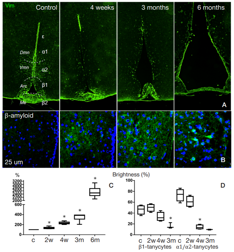
Fig. 1. Streptozotocin-induced changes in the third ventricle and adjacent structures. А. Identification of vimentin (Vim). B. Identification of β-amyloid in hypothalamic arcuate nucleus. C. Changes in the area of the third ventricle, % of the value of control (c). D. Changes in vimentin staining intensity (brightness, % of maximum levels) in the zone of β1 tanycyte and α1/α2 tanycyte localization. c — control; 2w, 4w, 3m, 6m — groups 2 and 4 weeks, 3 and 6 months after administration of streptozotocin; * — p < 0.05; populations of tanycytes (α1, α2, β1, β2) and ependymocytes (ε) are denoted by Greek letters; Dmn — dorsomedial hypothalamic nucleus; Vmn — ventromedial hypothalamic nucleus; Arc — hypothalamic arcuate nucleus; Ме — hypothalamic median eminence
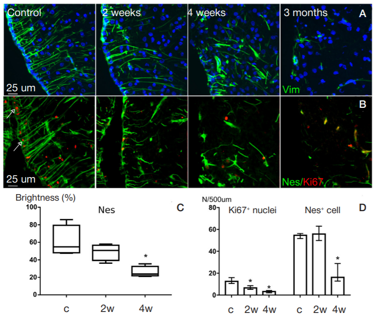
Fig. 2. Streptozotocin-induced changes in the morphology of tanycytes. A. Identification of vimentin (shown in green), DAPI staining of nuclei (blue). B. Identification of nestin (green) and Ki67 (red; arrows point to Ki67-positive nuclei). C. Changes in nestin staining intensity (brightness, % of maximum levels). D. Changes in the number of Nes+ and Ki67+ glial ependymal cells (number per unit of ventricular wall length). For notation see Fig. 1
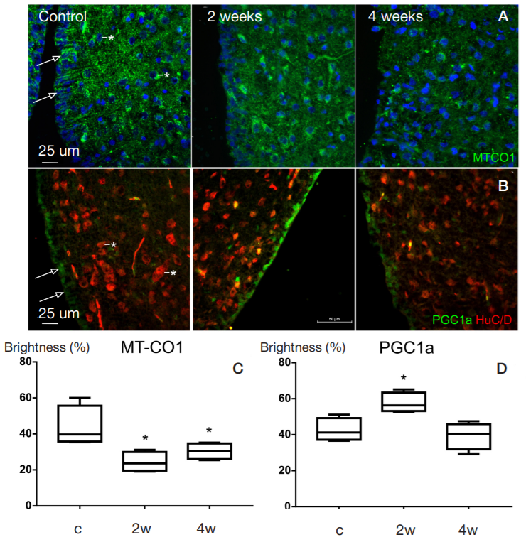
Fig. 3. Identification of tanycyte proteins associated with mitochondrial functions. А. Identification of MT-CO1 (mitochondrial respiratory chain complex IV); DAPI staining of nuclei (shown in blue). B. Identification of PGC1a (green); neurons (red) were identified using the anti-HuC/D antibodies. C. Changes in tanycyte cell body MT-CO1 staining intensity (brightness, % of maximum levels). D. Changes in tanycyte cell nuclei PGC1a staining intensity (brightness, % of maximum levels). Arrows point to tanycytes, neurons are marked with asterisks. For notation see Fig. 1
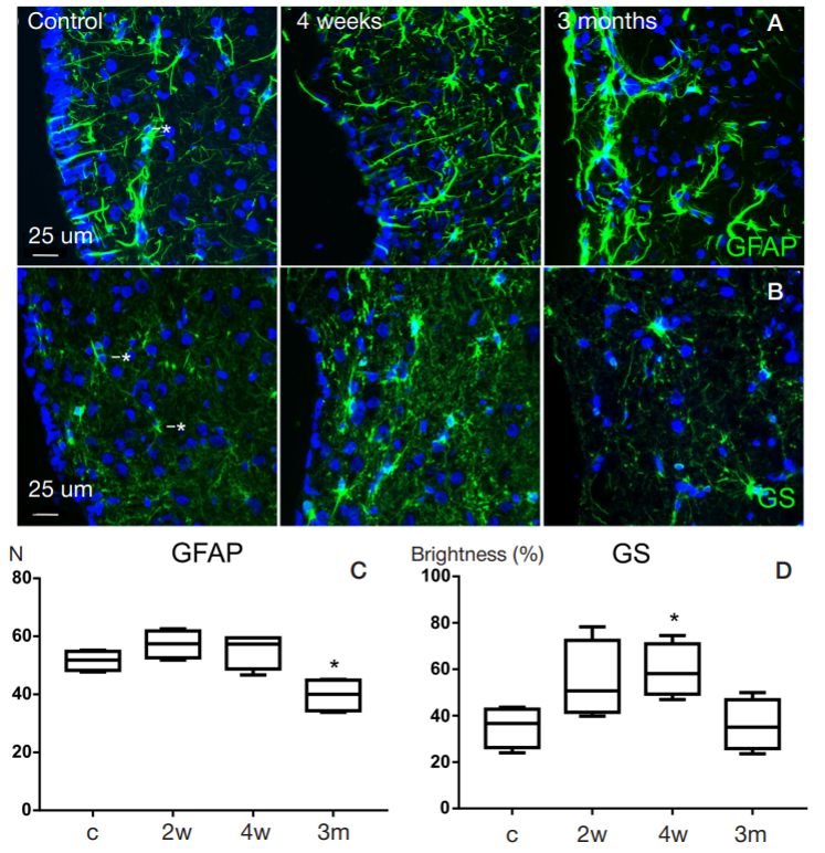
Fig. 4. Streptozotocin-induced changes in astroglia in the zone of hypothalamic arcuate nucleus (in the vicinity of the wall of the third ventricle). А. Identification of GFAP (shown in green); DAPI staining of nuclei (blue). B. Identification of glutamine synthetase (GS, green); DAPI staining of nuclei (blue). C. Changes in the number of GFAP-positive cells in the field of view. D. Changes in glutamine synthetase staining intensity (brightness, % of maximum levels). Astrocytes are marked with asterisks. For notation see Fig. 1
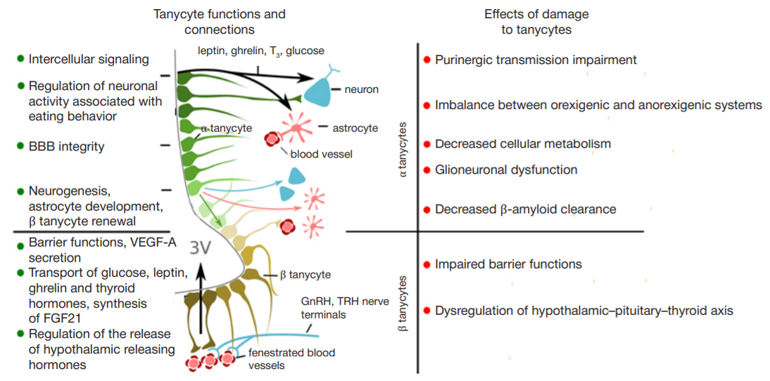
Fig. 5. Interaction between tanycytes and hypothalamic structures, and potential effects of damage to tanycytes. BBB — blood-brain barrier; VEGF-A — vascular endothelial growth factor A; FGF21 — fibroblast