ORIGINAL RESEARCH

Microglia and putative macrophages of the subfornical organ: structural and functional features

About authors

1 Institute of Experimental Medicine, St Petersburg, Russia

2 St Petersburg State University, St Petersburg, Russia

Correspondence should be addressed: Valeria V. Guselnikova
Akad. Pavlova, 12, St Petersburg, 197376, Russia; ur.xednay@aiirelav.avocinlesug

About paper

Funding: the study was supported by Russian Science Foundation, RSF Project № 22-25-00105, https://rscf.ru/project/22-25-00105/

Author contribution: Guselnikova VV — literature analysis, interpretation of the results, manuscript preparation; Razenkova VA — fluorescence immunoassay protocols development, confocal laser microscopy; Sufieva DA — histological processing, immunochemical staining, light microscopy; Korzhevskii DE — concept and planning of the study, editing of the manuscript.

Compliance with ethical standards: the study was approved by Ethical Review Board at the Institute of Experimental Medicine (Protocol № 1/22 of 18 February 2022) and carried out in full compliance with the 2013 Declaration of Helsinki.
Received: 2022-03-27 Accepted: 2022-04-18 Published online: 2022-04-28
|
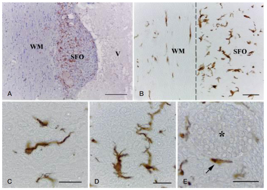
Fig. 1. Iba1-immunopositive cells in rat subfornical organ and adjacent white matter. A. Overall view. B. The boundary between subfornical organ and the underlying white matter. CE. Different morphotypes of Iba1-immunopositive cells within subfornical organ. SFO — subfornical organ, WM — white matter, V — third ventricle of the brain (cavity); the dashed line indicates the boundary between subfornical organ and white matter; the asterisk indicates blood vessel (lumen); the arrow indicates a small perivascular cell with few processes, containing Iba1. Scale bars, 200 µm (A), 50 µm (B), and 20 µm (CE)
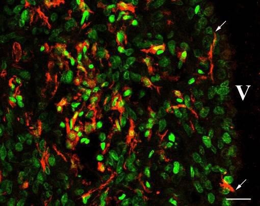
Fig. 2. Immunofluorescent detection of Iba1-positive cells in rat subfornical organ. The Iba1 specific signal is red (Cy3 fluorochrome) and the nuclei are green (SYTOX Green). V — third ventricle of the brain (cavity); the arrow indicates processes of the Iba1-containing cells reaching the ventricular cavity. Scale bar, 20 µm
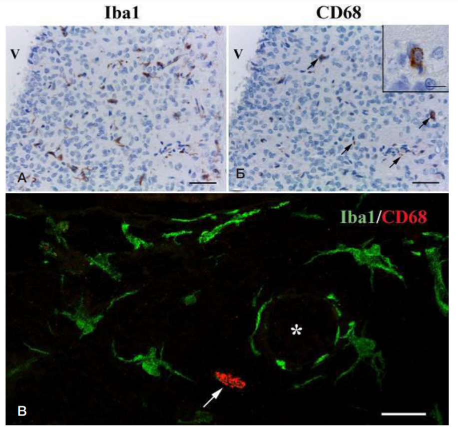
Fig. 3. Characteristic patterns of Iba1 and CD68 immunostaining in rat subfornical organ. A. Immunohistochemical reaction for Iba1 with alum hematoxylin nuclear counterstaining. B. Immunohistochemical reaction for CD68 with alum hematoxylin nuclear counterstaining; the arrows indicate CD68-immunopositive structures within the subfornical organ; the insert shows a magnified area comprising CD68-immunopositive cell. Images A and B represent histologically identical serial sections of the same specimen (light microscopy). C. Double-fluorescent immunostaining of Iba1/CD68 (confocal laser microscopy). The Iba1 signal is green (Cy2 fluorochrome) and the CD68 signal is red (Cy3 fluorochrome); the arrow indicates a CD68-containing cell; the asterisk indicates blood vessel (lumen). Scale bars, 50 µm (A, B) and 10 µm (B insert, C)