ORIGINAL RESEARCH

Comparative efficiency of accessible transfection methods in model cell lines for biotechnological applications

About authors

1 Engelhardt Institute of Molecular Biology, Moscow, Russia

2 Pirogov Russian National Research Medical University, Moscow, Russia

Correspondence should be addressed: Anastasia V. Lipatova
Vavilova, 32/1, Moscow, 119991, Russia; moc.liamg@vnaavotapil

About paper

Funding: the project was supported by the Russian Science Foundation (grant number 20-75-10157 of August 14, 2020 "Research on the possibilities of obtaining recombinant strains of oncolytic viruses with tumor-specific replication and immunomodulatory protein expression").

Author contribution: Vorobyev PO, Kochetkov DV, Vasilenko KV and Lipatova AV participated equally in the laboratory experiments, preparation of the figures and interpretation of the results.

Compliance with ethical standards: the study complies with the requirements of the World Medical Association Declaration of Helsinki.
Received: 2022-04-22 Accepted: 2022-05-30 Published online: 2022-06-23
|
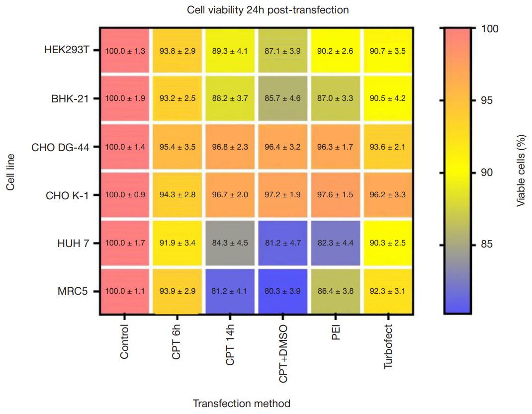
Fig. 1. The post-transfection MTT cell viability assay for PEI, TurboFect and CPT methods; Control — non-transfected cultures
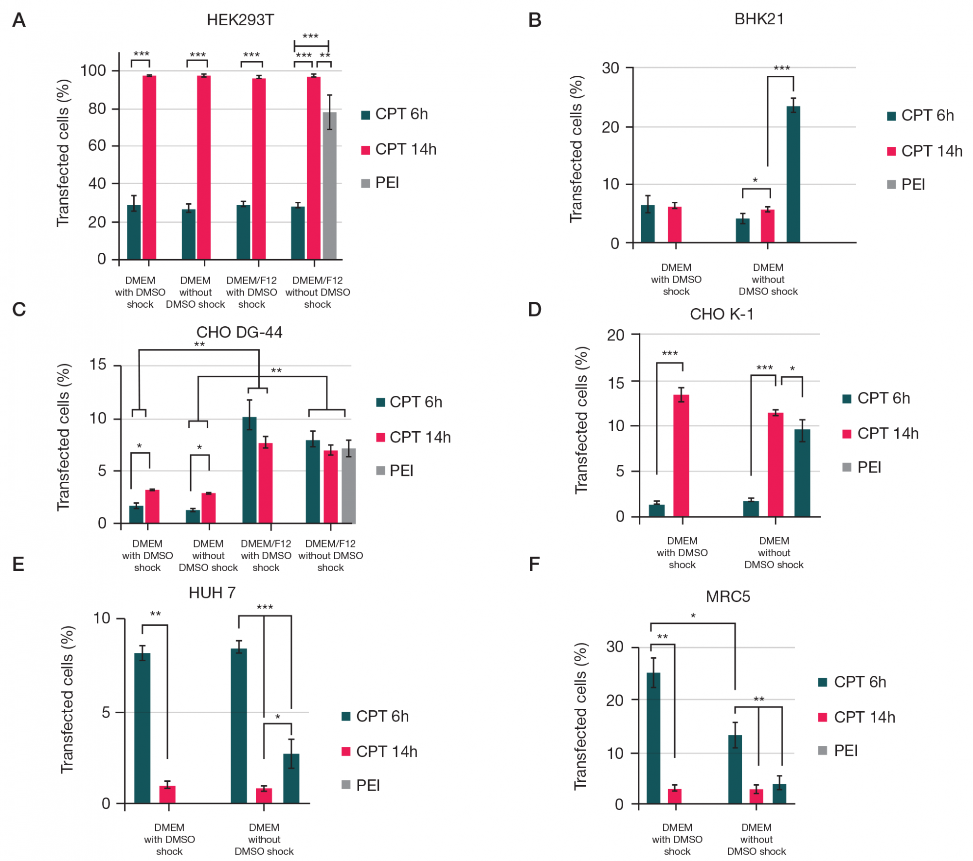
Fig. 2. Comparative efficiency of CPT and PEI monotransfections with Katushka expression plasmid (red fluorescence) in different model cell lines: HEK293T (A), BHK-21 (B), CHO DG-44 (C), CHO K-1 (D), Huh7 (E) and MRC5 (F). CPT 6 h — calcium phosphate transfection with 6 h incubation; CPT 14 h — calcium phosphate transfection with 14 h incubation
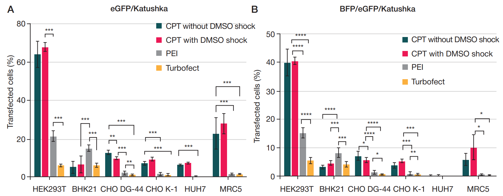
Fig. 3. Comparative efficiency of double cotransfections with eGFP and Katushka encoding plasmids (A) and triple cotransfections with BFP, eGFP and Katushka plasmids (B) in different model cell lines
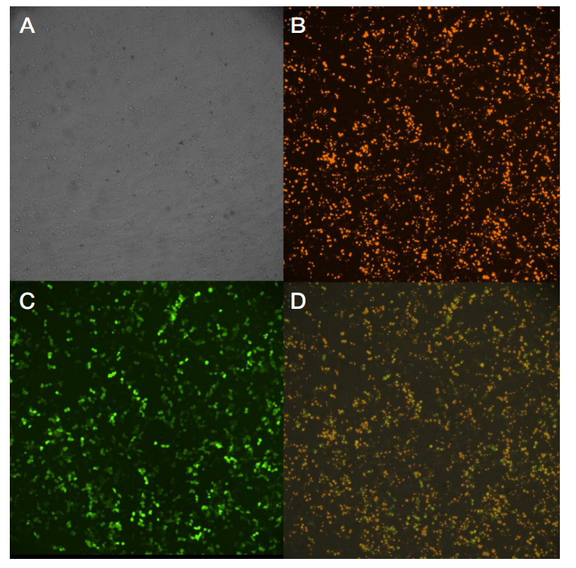
Fig. 4. Microphotographs of cell cultures cotransfected with genetic constructs encoding eGFP and Katushka fluorescent proteins. A. Light field. B. Katushka. C. eGFP. D. Merged image (magnification ×40)
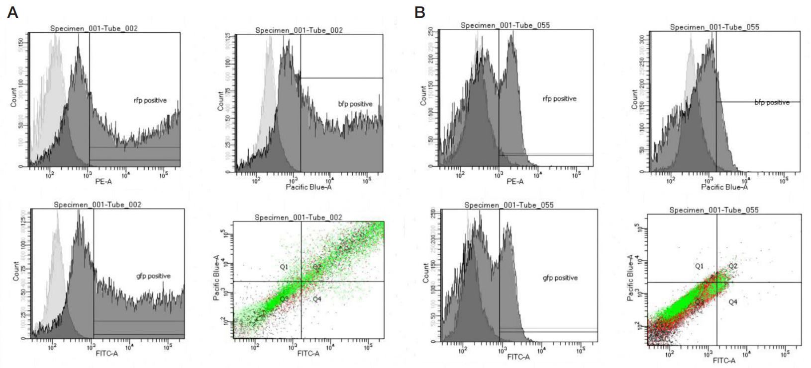
Fig. 5. Flow cytometry data for HEK293T and Huh7 cell lines (respectively, A and B); light contour — non-transfected control, dark contour — CPT. Populations single positive for particular fluorescent proteins (eGFP, Katushka or BFP) or triple-positive (Q2) are indicated
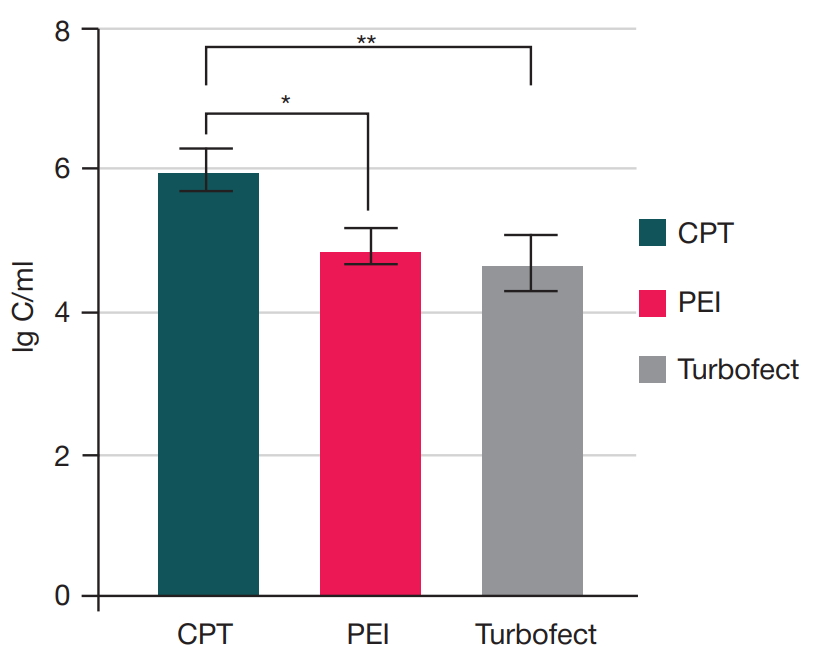
Fig. 6. Concentrations of lentiviral stocks produced with HEK293T cells using different transfection methods
Table 1. The 2× hepes buffer saline (HBS) composition
Table 2. Comparative efficiency of double cotransfections with eGFP and Katushka plasmids, measured 48 h post-transfection (best results in each row are highlighted)
Table 3. Comparative efficiency of triple cotransfections with BFP, eGFP and Katushka encoding plasmids, measured 48 h post-transfection (best results in each row are highlighted)
Note: ns — not significant.