METHOD

Foveal microsurgical reconstruction technique for macular hole

About authors

Pirogov Russian National Research Medical University, Moscow, Russia

Correspondence should be addressed: Khristo P. Takhchidi
Volokolamskoe shosse, 30, korp. 2, Moscow, 123182, Russia; moc.liamg@1031tph

About paper

Compliance with ethical standards: the study was approved by the Ethics Committee of the Pirogov Russian National Research Medical University (protocol № 224 dated 19 December 2022). All patients submitted the informed consent to surgical treatment and personal data processing.
Received: 2023-12-04 Accepted: 2023-12-18 Published online: 2023-12-31
|
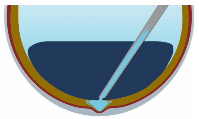
Fig. 1. Phases of the technique: vitreous cavity volume substitution with the perfluorocarbon liquid with subsequent aspiration of intraocular fluid from the hole area
Fig. 2. Phases of the technique: under exposure to negative pressure in the hole area, the aggregate foveal tissue of the macular hole fused walls is held in this position due to the perfluorocarbon liquid gravity
Fig. 3. Phases of the technique: application of the ACP prepared to the surface of the macular area of the retina with subsequent emergence of the hermetic plaque sealing the hole area (А) and filling the entire intrawound space (B)
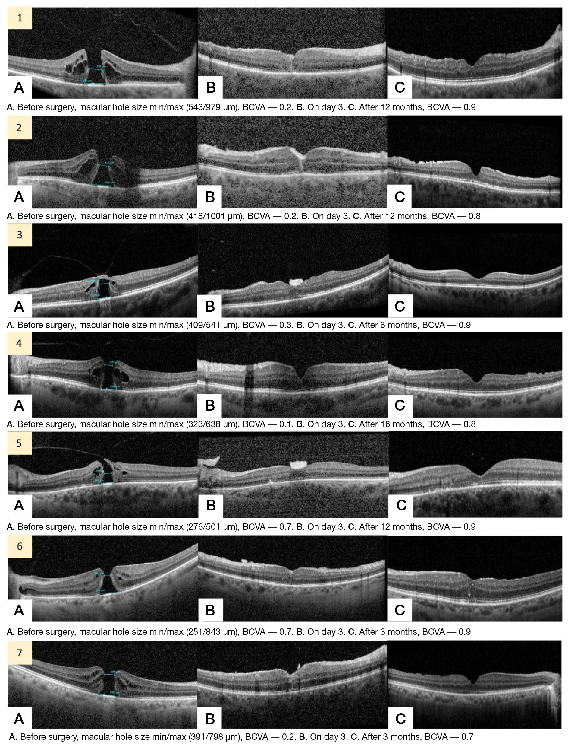
Fig. 4. OKT on the retina. Phases of macular hole closure after vitrectomy involving the use of ACP. А. Defect of all neuroretinal layers (macular hole). B. Follow up in the early postoperative period (on day 3). C. Follow up in the late postoperative period
Fig. 5. Phases of macular hole closure after vitrectomy involving the use of ACP. А. Defect of all neuroretinal layers (macular hole). B. Follow up in the early postoperative period (after 6 h). C. Follow up in the late postoperative period