ORIGINAL RESEARCH

LoRI, a new recombinant RNase inhibitor for in vitro applications

Sukhov DA1,2,3, Kholoshenko IV1,4, Petrova TV1, Romanenko GA1,2, Myshkin MYu3, Kost VYu3, Trofimov DYu1, Usman NYu5, Barsova EV3,5
About authors

1 DNA-Technology LLC, 117587, Moscow, Russia

2 MIREA — Russian Technological University, Moscow, Russia

3 Shemyakin–Ovchinnikov Institute of Bioorganic Chemistry RAS, Moscow, Russia

4 Bauman Moscow State Technical University, Moscow, Russia

5 Pirogov Russian National Research Medical University, Moscow, Russia

Correspondence should be addressed: Natalia Yu. Usman
Ostrovityanova 1/1, Moscow, 117997, Russia; ur.relbmar@namsu_n

About paper

Funding: The study was funded by DNA-Technology LLC.

Author contribution: Sukhov DA, Romanenko GA — protein engineering, expression and purification; Kholoshenko IV, Petrova TV — product characterization, writing; Myshkin MYu — protein structure analysis; Kost VYu — study design; Trofimov DYu — project supervision; Usman NYu — writing; Barsova EV — project coordination, writing.
Received: 2024-09-02 Accepted: 2024-10-09 Published online: 2024-10-28
|
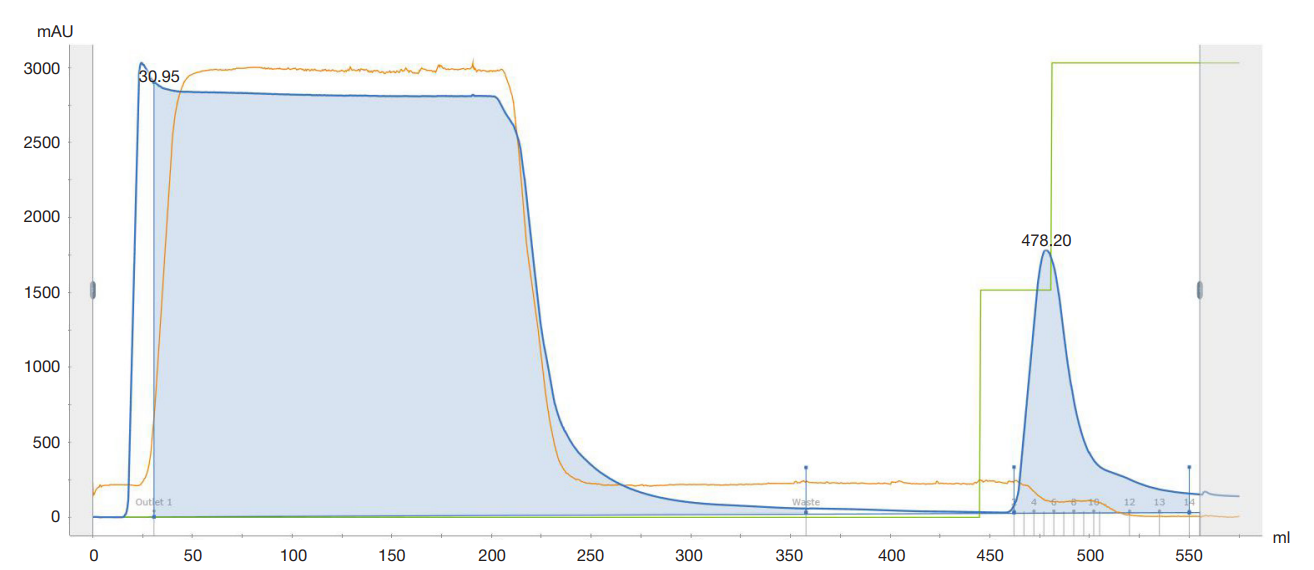
Fig. 1. Chromatography I profile (IMAC on Ni-INDIGO, Cube Biotech)
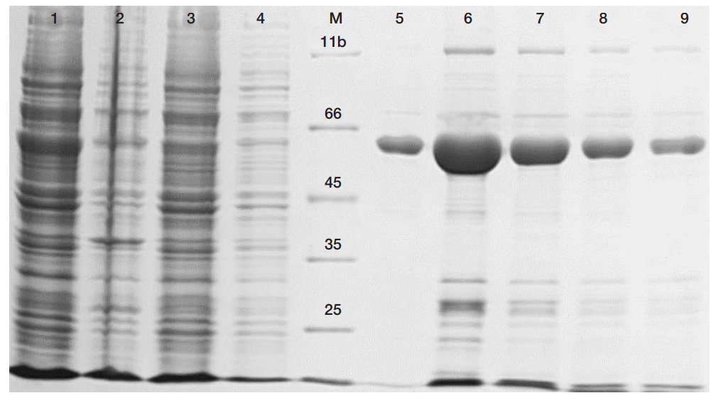
Fig. 2. Chromatography I samples, 10% SDS–PAGE. Lanes: 1 — supernatant; 2 — insoluble residue; 3 — flow-through; 4 — wash; M — protein molecular weight marker with band values, kDa, indicated in the image; 5–9 — eluted fractions 2, 5, 8, 10 and 11, respectively
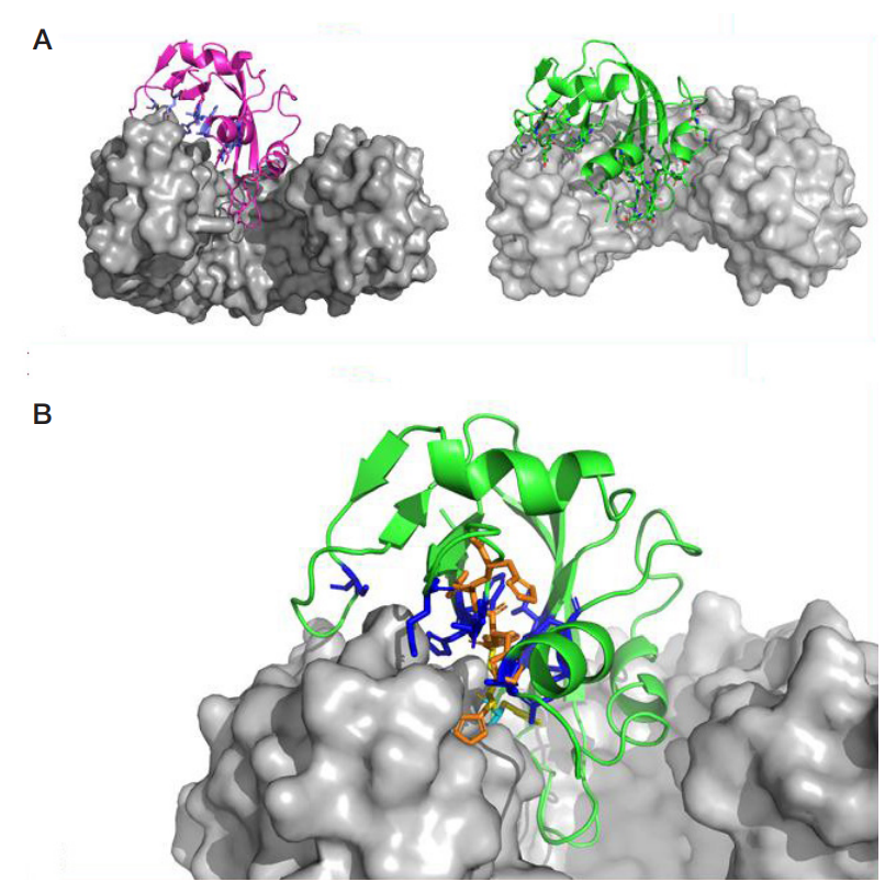
Fig. 3. Ribonuclease A (RibA) interacts with core inhibitor Rnh1 (grey surface). A. The enzyme contacts the inhibitor with its ligand-binding groove; the residues forming the active center are highlighted (PDB id: 1DFJ) [10]. B. Enlarged image of the C-terminus in close contact with residues of the active site in ribonuclease (blue) which sterically hinders the RNA binding. The histidine tag at the C-terminus (orange) disrupts the interaction by ‘protruding’ into RibA; overlay of AlphaFold3 model of Rnh1 with C-terminal His tag and crystal structure of Rnh1/RibA complex (PDB id: 1DFJ).
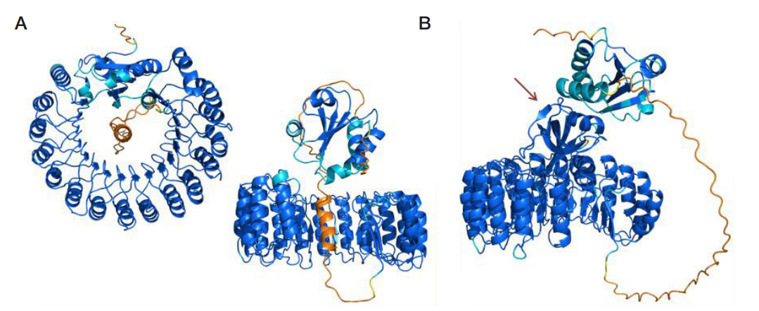
Fig. 4. AlphaFold3 models of Trx::Rnh1 fusion product. A. The product, native and unbound, comprising the horseshoe-shaped core inhibitor connected via linker to the compact Trx. The color scheme reflects predicted local distance difference test (pLDDT), a per-residue model confidence score provided by AlphaFold3, with pLDDT > 90, blue, high-confidence model; pLDDT 70–90, light-blue, high confidence for the backbone; pLDDT 50–70, yellow (low-confidence representation of a volatile structure); and pLDDT < 50, orange, region unstructured in isolation. Accordingly, the Rnh1 and Trx functional modules are imaged with high accuracy, whereas representation of the linker is conditional: it can be oriented and located differently e.g. outside the horseshoe. B. RibA (arrow) appears sandwiched between Trx and Rnh1 modules of the fusion product
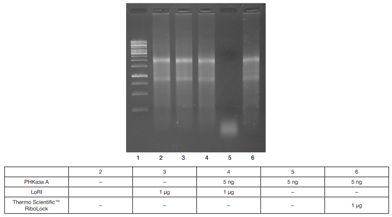
Fig. 5. RNA stability assay, pilot series. Lane 1: Thermo Scientific™ GeneRuler 1kb DNA Ladder. Lanes 2–6: 1 µg RNA +. The treatment proceeded at 37 °C for 30 min.
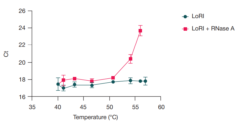
Fig. 6. Reverse transcription PCR data for RNA stability assay at 40–57 °C using 1 µg RNA + 2 µg LoRI with or without RNase A (2.5 ng), treatment time — 30 min
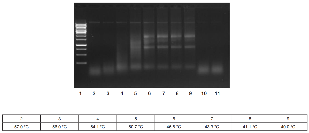
Fig. 7. RNA stability assay at 40–57 °C, treatment time— 30 min. Lane 1: Thermo Scientific™ GeneRuler 1kb DNA Ladder Lanes 2–9: 1 µg RNA + 2.5 ng RNase A + 2 µg LoRI at
Lanes 10–11: 1 µg RNA + 2.5 ng RNase A, no inhibitor added, incubated at 57.0 and 40.0 °C, respectively