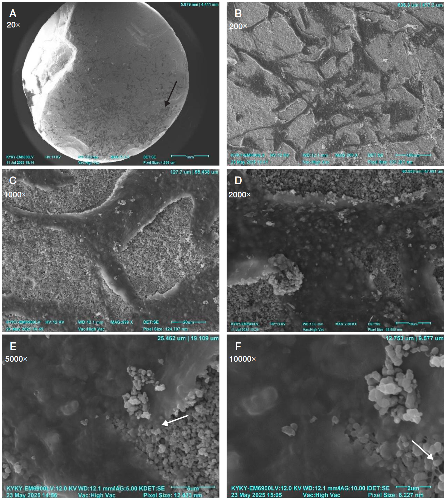
Fig. 1. Scans of the surface of tricalcium phosphate samples after sintering at 800 °C with growing mesenchymal stem cells from adipose tissue, 7 days of cultivation. The black arrow shows dark cells on the surface of a cylindrical tricalcium phosphate sample, and the white arrow shows thin extensions of the membrane. The cells are large, form a cluster, are strongly spread out, in contact with each other by wide extensions; the the cell surface is smooth, there are thin extensions of the membrane visible under the cell body and along the periphery. Magnification from 20× to 10000×
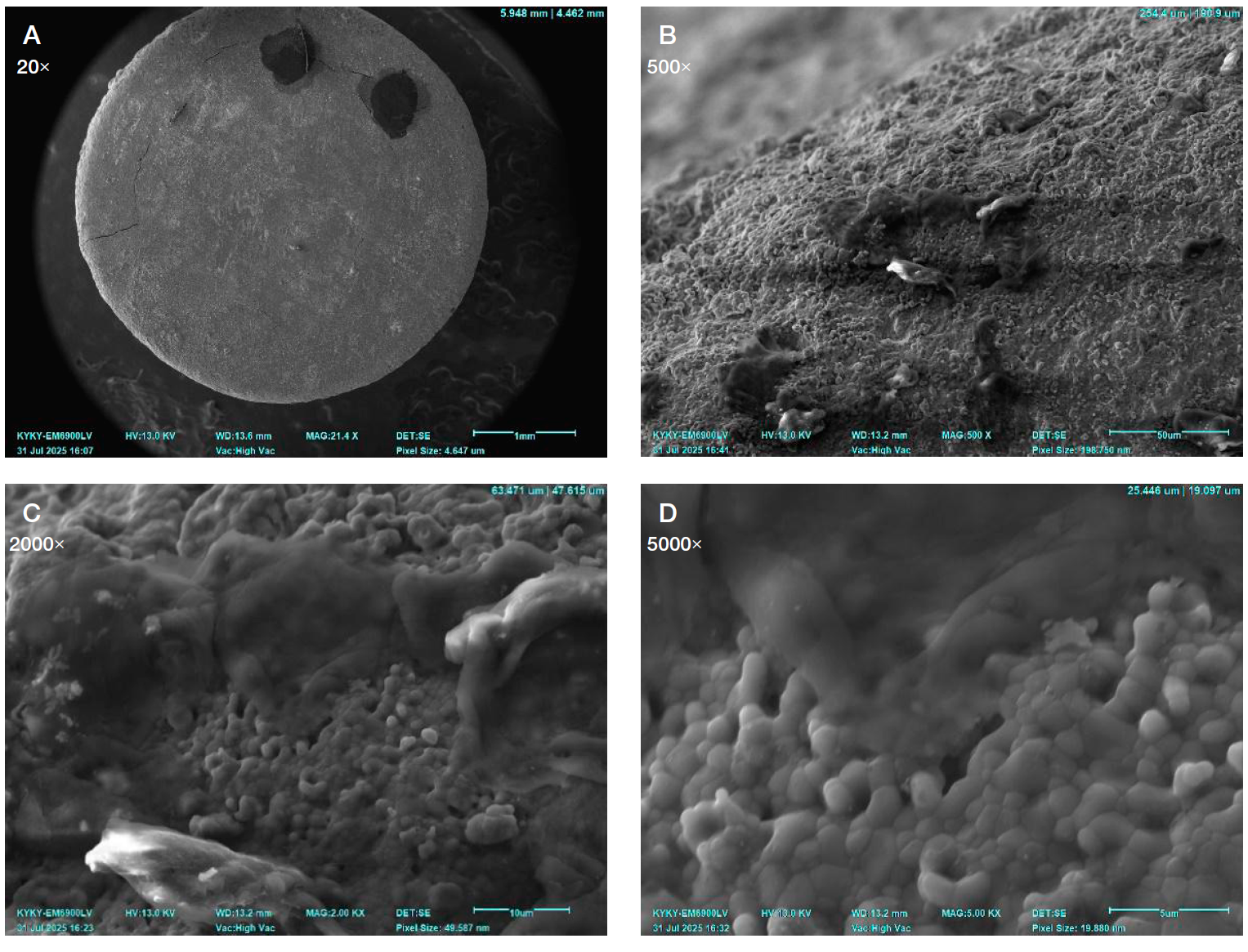
Fig. 2. Scans of the surface of hydroxyapatite samples after sintering at 1250 °C with growing mesenchymal stem cells from adipose tissue, 7 days of cultivation. The surface of the cells is folded, smooth, the cell body is creeping. Magnification from 20× to 5000×
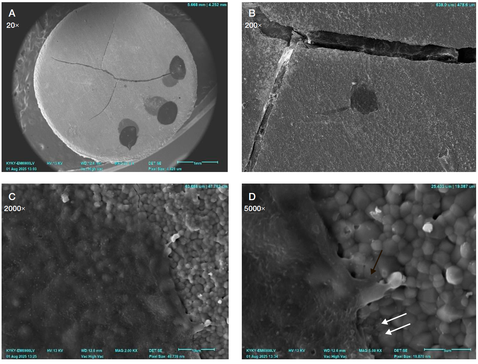
Fig. 3. Scans of the surface of tricalcium phosphate samples after sintering at 1250 °C with growing mesenchymal stem cells from adipose tissue, 7 days of cultivation. The black arrow point to a wide pseudopod. The white arrow — to a thin extension that widens at the end. Magnification 20x–5000x. The cell has a high degree of adhesion, it fits firmly to the surface, is strongly flattened; underneath, the contour of the material granules are visible, the surface has small depressions, extensions of various thicknesses are seen along the perimeter of the cell and are visible under its body
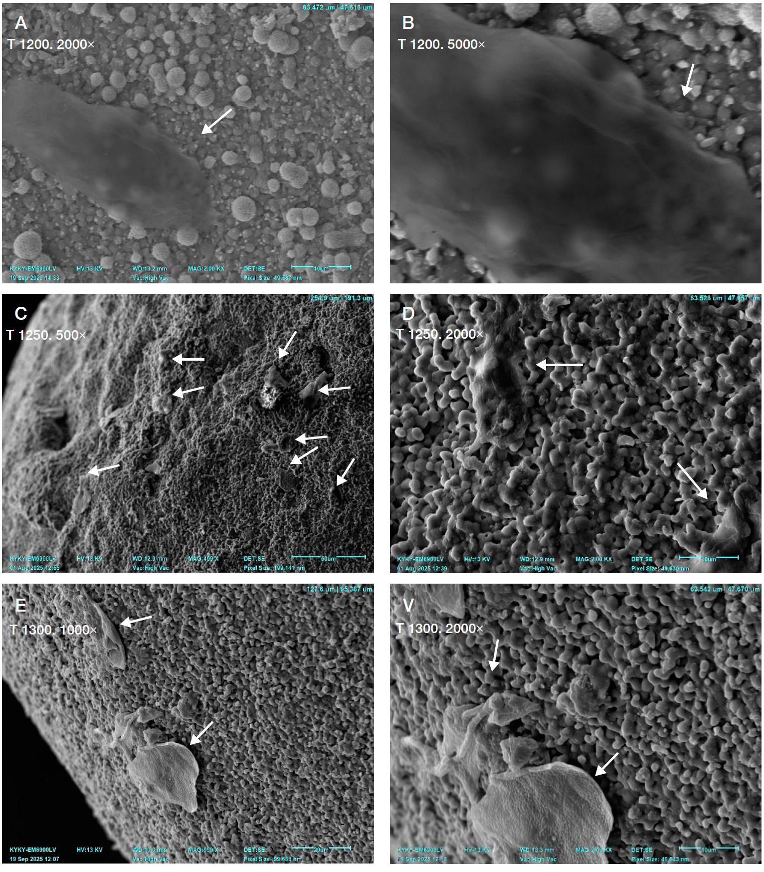
Fig. 4. The nature of cell growth on bone allograft samples with different physical processing parameters. Scans. T1200 — sintering temperature of 1200 ° C, T1250 — sintering temperature of 1250 °C, T1300 — sintering temperature of 1300 °C. The degree of cell adhesion to the material is different. The white arrows point to the cells on the surface of the allograft material. A, B. Maximum adhesion: the cells adhere tightly to the surface, they are strongly flattened, the contour of the granules of the material is visible under the cell body, the surface is smooth, with faintly noticeable folds, there are extensions of varying thicknesses (mostly thin, around the perimeter of the cell, visible under its body). C, D. Moderate adhesion: the cells adhere to the surface, in some areas the edges of the cell are raised, the surface has clearly visible folds, and the extensions are predominantly wide. E, F. Low adhesion: the cells are slightly spread out, the edges of the cells are noticeably raised around the perimeter, the cell surface has small bumps, there are no extensions
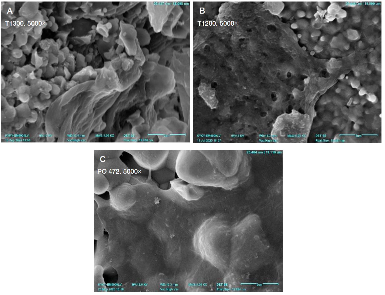
Fig. 5. Morphological features of mesenchymal stem cells from adipose tissue on the surface of tricalcium phosphate samples. Scans. A. Folded surface of the cell, smooth, low degree of spreading, adhesion at individual points, no extensions of the cell body. B. The cell is spread out, almost completely adhered, the surface is bumpy, there are several wide and separate thin extensions along the periphery. C. The cell is strongly spread out, fully adhered, the surface is smooth, there are many wide and thin extensions under the cell body and along the periphery. T1200, T1300 — the sintering temperature of the material during the manufacture of samples in °C. RO 472 — laboratory sample marking

