Copyright: © 2025 by the authors. Licensee: Pirogov University.
This article is an open access article distributed under the terms and conditions of the Creative Commons Attribution license (CC BY).

ORIGINAL RESEARCH

Antiviral activity of mRNAS encoding intracellular scFv antibodies against conserved influenza virus epitopes

Plotnikova MA , Oleynik VA , Klotchenko SA
About authors

Smorodintsev Research Institute of Influenza, St. Petersburg, Russia

Correspondence should be addressed: Sergey A. Klotchenko
Professora Popova, 15/17, Saint Petersburg, 197022, Russia; ur.liam@kitafsof

About paper

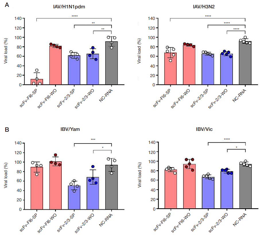
Funding: This study was supported by financially by the Russian Science Foundation, Agreement No. 24-25-00488: "Study of the antiviral potential of intracellular scFv antibodies against influenza virus" (supervisor — S.A. Klotchenko), https://rscf.ru/project/24-25-00488/

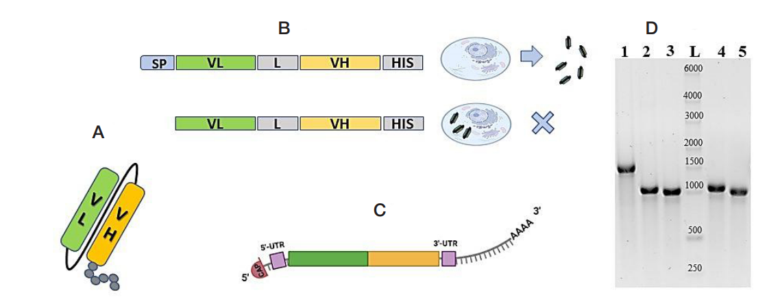
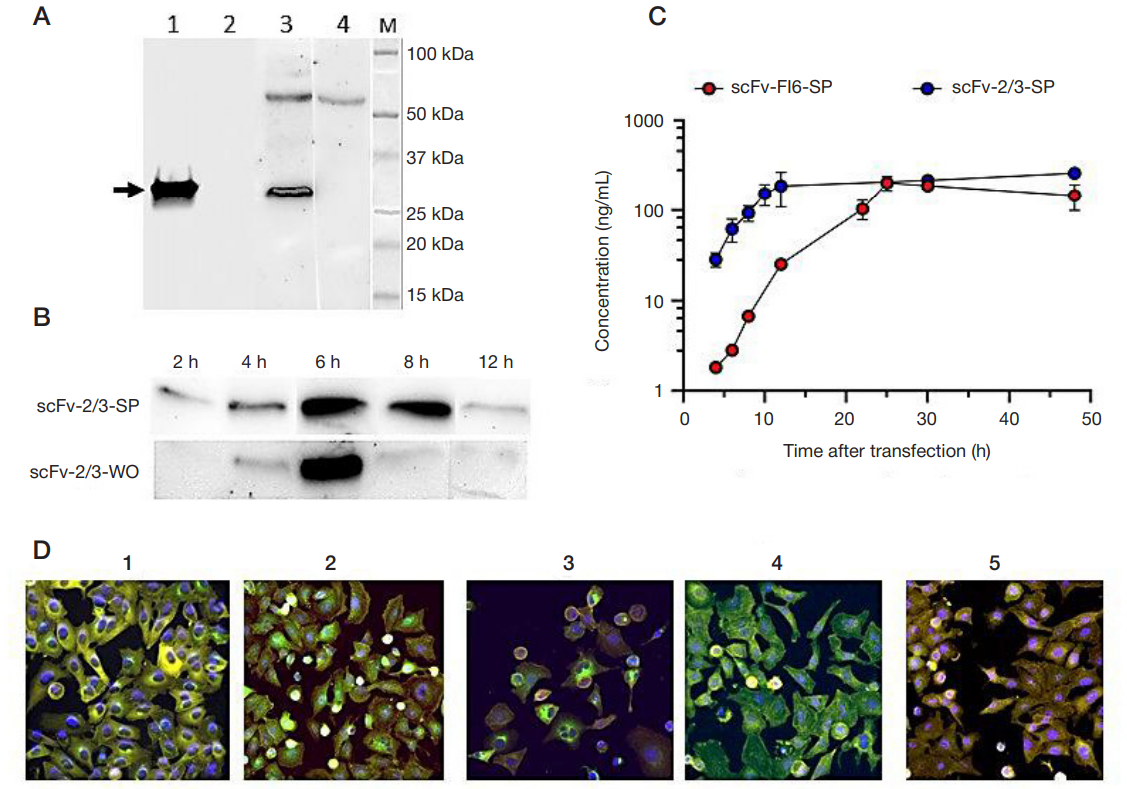
Author contribution: Plotnikova MA — design of structures, conducting experiments, registration and analysis of results, statistical processing, manuscript authoring and formatting; Oleynik VA — conducting experiments, registration of results; Klotchenko SA — study design, preparation and characterization of mRNA preparations, conducting experiments, registration and analysis of results, manuscript editing.

Received: 2025-11-27 Accepted: 2025-12-10 Published online: 2025-12-26
|
Fig. 1. Basic structure of the developed scFv antibody fragments against influenza A and B viruses and the exogenous mRNAs encoding them. A. Structure of scFvs composed of variable domains of the light (green) and heavy (orange) chains. The black line indicates the linker peptide (G4S)4. Gray circles indicate the C-terminal 6×His-tag sequence. B. Structure of the coding regions of secreted and cytosolic scFv antibodies. The key difference is the presence of a signal peptide (SP) at the N terminus in the secreted variant. VL and VH denote the variable domains of the light and heavy chains, respectively; L, the linker peptide; HIS, the 6'His tag sequence. C. Structure of the exogenous mRNA used for intracellular scFv translation. The 5' cap (m7GmAmG) is shown at the 5' terminus; the 5' UTR and 3'UTR regulatory regions are shown in lilac; green and yellow blocks represent the scFv coding region; the extended gray line with notches represents the poly(A) tail. D. Agarose gel electrophoretic analysis of purified exogenous mRNA preparations: lane 1, negative control mRNA encoding the heavy chain of an immunoglobulin against an irrelevant antigen (NC-RNA), estimated length 1792 nt; lane 2, mRNA encoding scFv-FI6-SP, 1219 nt; lane 3, mRNA encoding scFv-FI6-WO, 1162 nt; lane 4, mRNA encoding scFv-2/3-SP, 1201 nt; lane 5, mRNA encoding scFv-2/3-WO, 1138 nt. L, RNA molecular weight marker (RiboRuler High Range RNA Ladder; Thermo Fisher Scientific, USA). Marker sizes (nt) are indicated on the lane
Fig. 2. Translational activity of exogenous mRNAs encoding cytosolic (WO) and secreted (SP) forms of scFv antibody fragments against influenza A and B viruses. A. Western blot analysis of scFv-2/3 antibody fragments in culture medium and MDCK cell lysates using antibodies specific to the 6'His tag sequence. Similar results were obtained for scFv-FI6 but are not shown. Lanes 1 and 2 show culture medium from MDCK cells transfected with mRNAs encoding scFv-2/3-SP and scFv-2/3WO, respectively; lanes 3 and 4 show cell lysates from MDCK cells transfected with mRNAs encoding scFv-2/3-SP and scFv-2/3-WO, respectively. M, molecular weight marker (Precision Plus Protein Kaleidoscope Preferred Protein Standards; Bio-Rad, USA); corresponding molecular weights (kDa) are indicated to the right. All samples were collected 24 h after mRNA cell transfection. B. Western blot analysis of scFv-2/3 antibody fragments in MDCK cell lysates at different time points using anti–6'His-tag antibodies. The upper panel shows intracellular production of scFv-2/3-SP, and the lower panel shows scFv-2/3-WO. Sampling times after mRNA transfection are indicated at the top (hours). C. ELISA analysis of scFv concentrations in culture medium after transfection of A549 cells with mRNAs encoding scFvFI6-SP (red) and scFv-2/3-SP (blue). Data points represent mean values from two biological replicates ± SD. D. Representative fluorescence microscopy images of fixed A549 cells transfected with exogenous mRNAs encoding: (1) scFv-FI6-SP, (2) scFv-FI6-WO, (3) scFv-2/3-SP, (4) scFv-2/3-WO, and (5) NC-RNA. The actin cytoskeleton (yellow) was visualized using rhodamine-conjugated phalloidin, and nuclei (blue) were stained with DAPI. scFv fragments (green) were detected using primary mouse monoclonal anti-6'His-tag antibodies followed by Alexa Fluor 488-conjugated goat anti-mouse secondary antibodies. Images were acquired and processed using a Cytell cell imaging system (40× objective); individual channels were pseudocolored in RGB during image processing. Enlarged views of the processed images are shown
Fig. 3. In-cell ELISA analysis of the antiviral activity of exogenous mRNAs encoding cytosolic (WO) and secreted (SP) forms of scFv antibody fragments against influenza A and B viruses. A. Intracellular levels of influenza A virus (IAV) after infection with A/California/07/09 (H1N1pdm) (left) and A/Cambodia/e0826360/2020 (H3N2) (right) strains. B. Intracellular levels of influenza B virus (IBV) after infection with B/Phuket/3073/13 (Yamagata) (left) and B/Malaysia/2506/2004 (Victoria) (right) strains. Data points represent mean values for four samples ± SD. Red bars indicate therapeutic administration of exogenous mRNAs encoding scFv fragments against IAV hemagglutinin; blue bars indicate administration of exogenous mRNAs encoding scFv fragments against IBV nucleoprotein. Gray bars show mean values for cells transfected with exogenous mRNA encoding the heavy chain of an immunoglobulin specific to an irrelevant antigen (negative control). Statistical significance was determined using one-way ANOVA followed by Holm-Šídák multiple comparison test. Asterisks indicate significant differences compared with the negative control (NC-RNA): * — pvalue < 0.0332; * * — pvalue < 0.0021; * * * — pvalue < 0.0002; * * * * — pvalue < 0.0001