Copyright: © 2026 by the authors. Licensee: Pirogov University.
This article is an open access article distributed under the terms and conditions of the Creative Commons Attribution license (CC BY).

ORIGINAL RESEARCH

Effects of standard and photoactivated platelet-rich plasma on human meniscus fibrochondrocytes in IL-1β-induced inflammation

About authors

1 MEDSI Clinic, Clinical Hospital No. 1, Moscow, Russia

2 Scientific and Diagnostic Center "Polyclinic on Smolenskaya", Moscow, Russia

3 St. Luke Lugansk State Medical University, Russia

Correspondence should be addressed: Aleksey S. Ivanov
A. Dikogo 16A, 73, 111396, Moscow, Russia, ur.liam@5891golotamvartahsas

About paper

Author contribution: Kerimov KB — study design, experimental procedure, calculations; Ivanov AS — study design, experimental design; Tananakina TP — study design, experimental design, scientific supervision; Kashchenko SA — scientific editing, scientific supervision, part in preparing the experiment; Pogorelova IA — technical proofreading, part in preparing the experiment.

Compliance with ethical standards: the study approved by the Ethics Committee of the St. Luke Lugansk State Medical University (protocol No. 2 dated May 26, 2026) was fully compliant with the Eurasian Economic Commission (EEC) Board "Guidelines for the Use of Laboratory (Experimental) Animals in Preclinical (Non-Clinical) Studies" dated November 14, 2023, No. 33, principles of the European Convention for the Protection of Vertebrate Animals used for Experimental and Other Scientific Purposes, as well as the Directive of the European Parliament and of the Council of the European Union on the Protection of Animals Used for Scientific Purposes. All the patients submitted the written informed consent for the material collected to be used for experimental purposes.
Received: 2026-06-27 Accepted: 2026-08-20 Published online: 2026-08-26
|
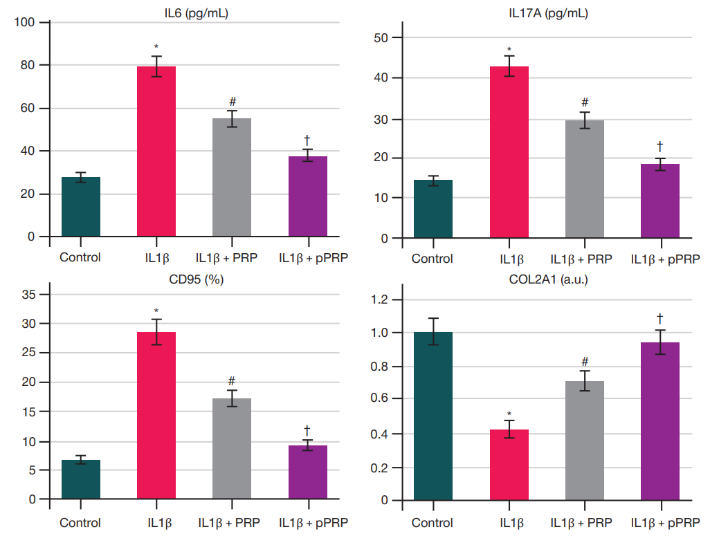
Fig. 1. Inflammatory (IL6, IL17A), phenotypic (CD95), and matrix (COL2A1) profiles of human meniscus chondrocytes in vitro I the IL1β-induced inflammation adjusted with standard and photoactivated PRP. Control — intact control group not exposed to IL1β; IL1β + 0,9% NaCl — group 2, IL1β + PRP — group 3; IL1β + p PRP — group 4. Each individual point on the graph corresponds to the average value of technical replicates for one biological donor out of 12. # — p < 0.05 compared to the “IL1β” group; † — p < 0.05 compared to the "IL1β + PRP” group (post-hoc Tukey’s test for repeated measurements)
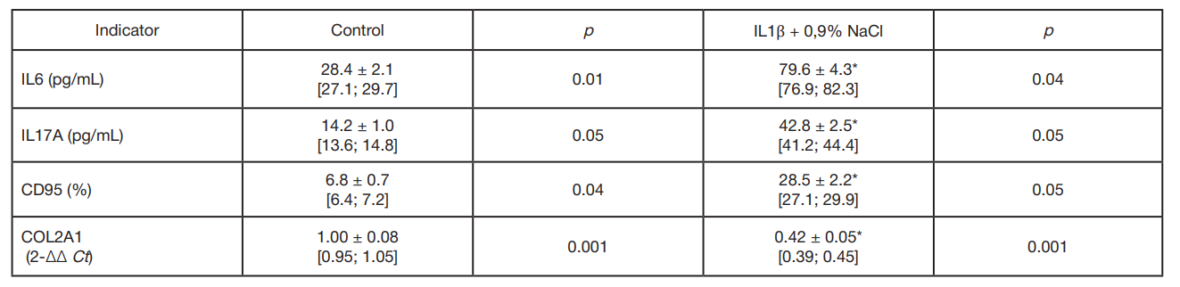
Table 1. Effects of interleukin-1β on the secretory, phenotypic and matrix profiled of human meniscus chondrocytes in vitro after the 48-h incubation (M ± SD [95% CI], n = 12)
Note: * — p < 0.05 compared to control.
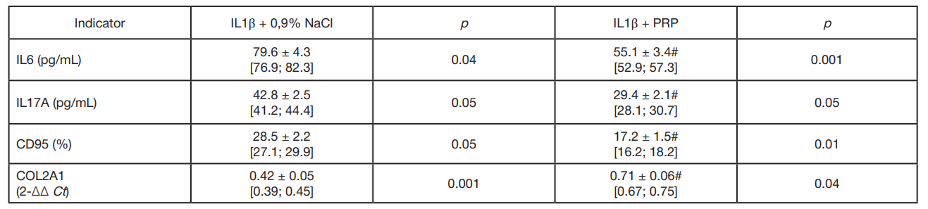
Table 2. Effects of platelet-rich plasma on the inflammatory, phenotypic, and matrix profiles of human meniscus chondrocytes in vitro in the context of interleukin-1β induced inflammation (M ± SD [95% CI], n = 12).
Note: # — p < 0.05 compared to the group of IL1β.
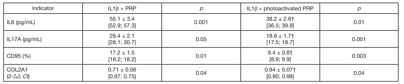
Table 3. Comparative analysis of the effects of standard and photoactivated platelet-rich plasma on the inflammatory, phenotypic, and matrix profiles of human meniscus chondrocytes in vitro in the context of interleukin-1β-induced inflammation (M ± SD [95% CI], n = 12).
Note: † — p < 0.05 compared to the group of standard PRP.