ISSN Print 2500–1094
ISSN Online 2542–1204
BIOMEDICAL JOURNAL OF PIROGOV UNIVERSITY (MOSCOW, RUSSIA)

1 Orekhovich Institute of Biomedical Chemistry, Moscow, Russia
2 RMA “Perspektiva”, Novosibirsk, Russia
Correspondence should be addressed: Alexander L. Rusanov
Pogodinskaya 10, bld. 8, Moscow, 119121; moc.liamg@vonasur.l.rednaxela
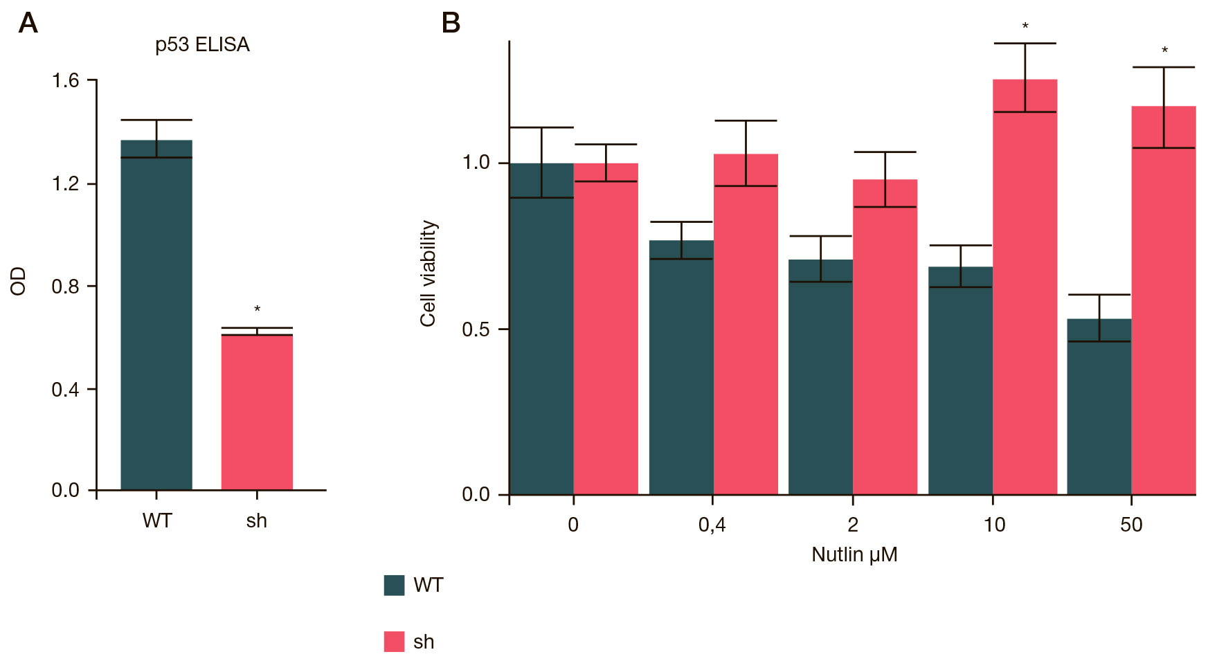
Funding: the study involving p53 gene knockdown, ELISA and PCR tests was performed as part of the Fundamental Scientific Research Programs of the State Academies of Sciences for 2013–2020; experiments with Nutlin-3a were carried out by RMA “Perspektiva” and supported by RFBR, project № 18-44-540031/19.
Author contribution: Luzgina NG, Rusanov AL — study concept; Romashin DD, Kozhin PM, Luzgina NG, Rusanov AL — study design and literature analysis; Romashin DD, Kozhin PM, Karagyaur MN — study planning and execution; Kozhin PM, Romashin DD, Luzgina NG, Rusanov AL — data analysis and interpretation; Kozhin PM, Romashin DD — manuscript writing; Kozhin PM, Romashin DD, Karagyaur MN, Luzgina NG, Rusanov AL — manuscript editing, preparation of the final version of the article.
Compliance with ethical standards: the study was carried out in accordance with the World Medical Association Declaration of Helsinki.