ORIGINAL RESEARCH

Experimental assessment of biological potential of collagen membranes in reconstruction of full-thickness hyaline cartilage defects

Lazishvili GD1, Egiazaryan KA1, Nikishin DV2, Voroncov AA3, Klinov DV4
About authors

1 Pirogov Russian National Research Medical University, Moscow, Russia

2 OOO Aptos Group, Moscow, Russia

3 Doctor Vorontsov's Veterinary Center for Surgery and Oncology, Moscow, Russia

4 Federal Research and Clinical Center of Physical-Chemical Medicine of Federal Medical Biological Agency, Moscow, Russia

Correspondence should be addressed: Guram D. Lazishvili
Ostrovityanova, 1, 117997, Moscow; moc.liamg@zalmarug

About paper

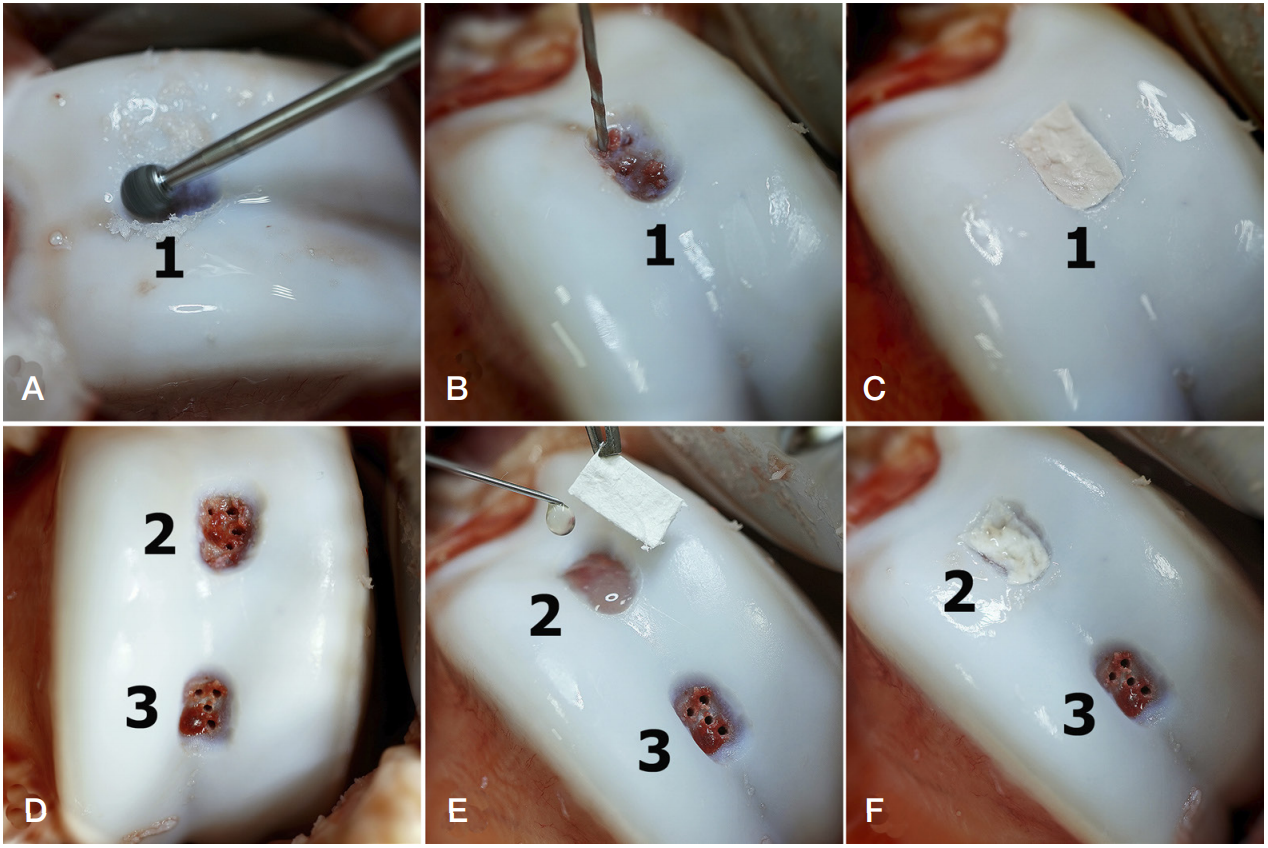
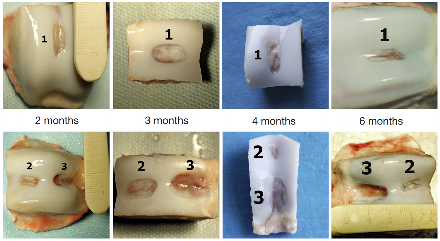
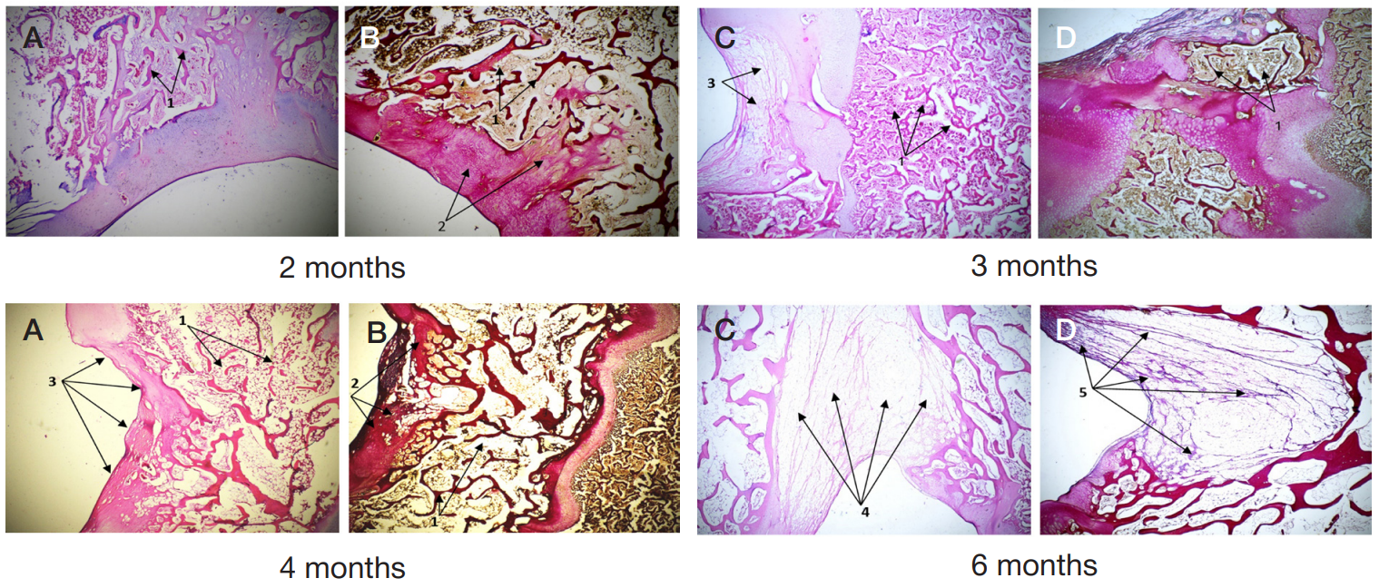
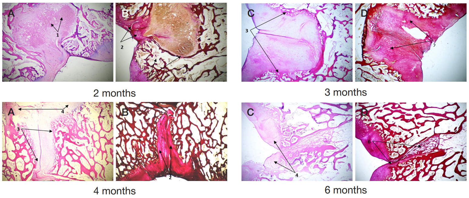
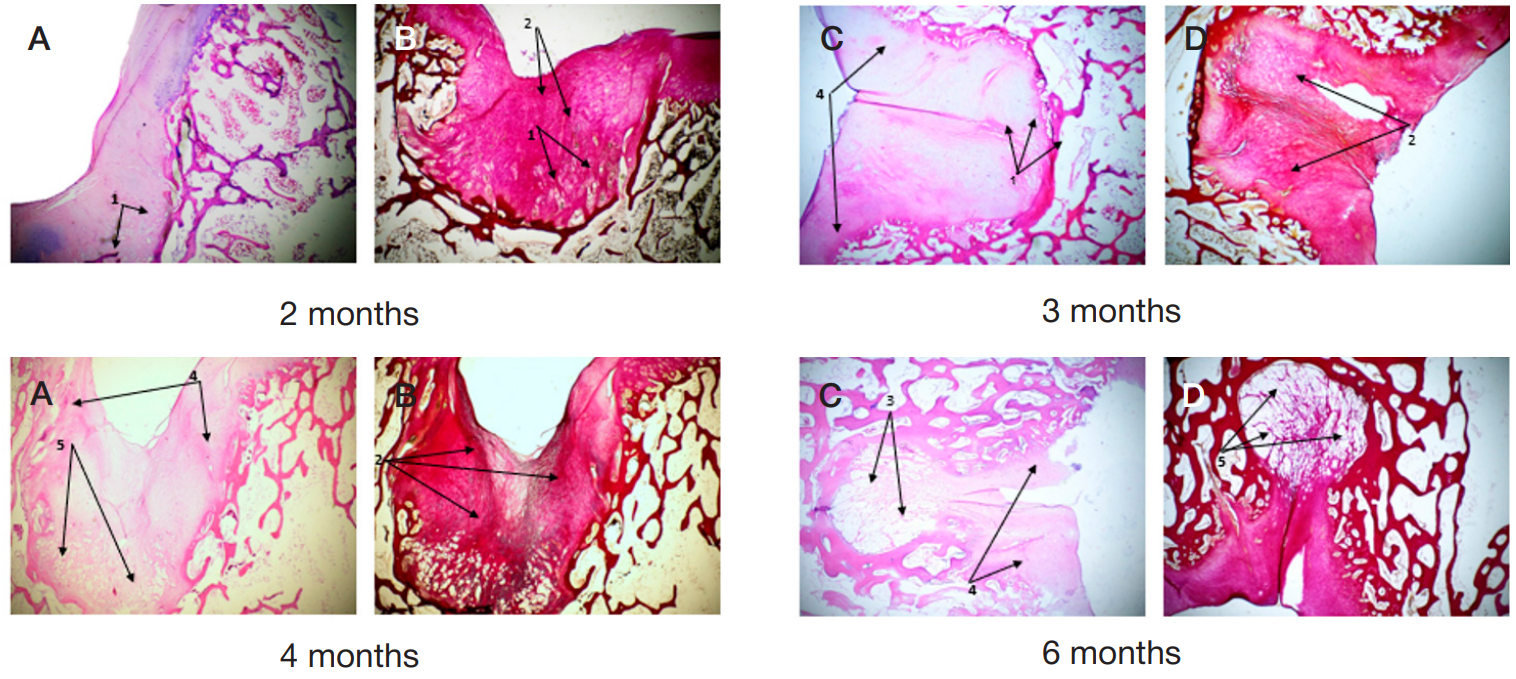
Author contribution: Lazishvili GD — design of the experiment, participation in the experimental surgery, analysis of literature and experimental materials, article authoring; Yeghiazaryan KA — analysis of literature, experimental results; Nikishin DV — processing and analysis of the experimental data, article authoring; Vorontsov AA — execution of the experimental surgery; Klinov DV — Ortokeep collagen membrane design and development.

Compliance with ethical standards: the study was approved by the Ethics Committee of the Center for Preclinical Research of Penza (Minutes № 1–19 of March 11, 2019). The animals were kept and used in compliance with the ethical standards and International requirements for humane treatment of laboratory (experimental) animals, as well as GOST R ISO 10993-1-2009 Medical Devices.

Received: 2021-07-19 Accepted: 2021-08-05 Published online: 2021-08-20
|
Fig. 1. Stages of formation of full-thickness defects and implantation of collagen membranes. 1 — defect № 1 (implantation of the Ortokeep membrane); 2 — defect № 2 (implantation of the Chondro-Gide membrane); 3 — defect № 3 (no membrane implantation)
Fig. 2. Macro specimen at various timepoints post surgery. 1 — defect № 1 after implantation of the Ortokeep membrane; 2 — defect № 2 after implantation of the Chondro-Gide membrane; 3 — defect № 3, no membrane implantation
Fig. 3. Microscopic examination of defect № 3, control group; staining with hematoxylin and eosin, — 40 (A); Van Gieson's stain, ×40 (B). 1 — osteodystrophy; 2 — coarse fibrous connective tissue; 3 — developing "crater" defect in cartilage and bone tissues; 4 — groups of adipocytes filling the "crater" defect; 5 — bundles of collagen fibers between islets of adipocytes
Fig. 4. Microscopic examination of defect № 1 after implantation of the Ortokeep membrane; staining with hematoxylin and eosin, ×40 (A); Van Gieson's stain, ×40 (B). 1 — fibrin; 2 — coarse fibrous connective tissue; 3 — developing cylindrical defect, active neoosteogenesis evident at the edges; 4 — neochondrogenesis; 5 — the defect shrunk sharply with formation of a slit cavity in the center
Fig. 5. Microscopic examination of defect № 2 after implantation of the Chondro-Gide membrane, stained with hematoxylin and eosin, ×40 (A); Van Gieson's stain, ×40 (B). 1 — fibrin; 2 — coarse fibrous connective tissue; 3 — emerging bulbous defect, active neoosteogenesis evident along the edges; 4 — neochondrogenesis; 5 — the bed of the bone defect filled with adipocytes
Table. Dimensional characteristics of the histological structure of the defect's center, various types of treatment