ORIGINAL RESEARCH

Single-domain antibody for binding the conserved epitope in the SARS-CoV-2 spike protein receptor-binding domain

Vorobyev PO1, Tillib SV1,2
About authors

1 Engelhardt Institute of Molecular Biology, Russian Academy of Sciences, Moscow, Russia

2 Institute of Gene Biology, Russian Academy of Sciences, Moscow, Russia

Correspondence should be addressed: Sergei V. Tillib
Vavilov str., 34/5, Moscow, 119334, Russia; ur.ygoloibeneg@billit moc.liamg@billit.iegres

About paper

Funding: the study was supported by the Ministry of Science and Higher Education of the Russian Federation (agreement № 075-15-2021-1086, contract № RF––193021X0015).

Acknowledgements: the authors thank M.V. Rutovskaya, Severtsov Institute of Ecology and Evolution of the Russian Academy of Sciences, for assistance in immunization of the camel.

Author contribution: Vorobyev PO — molecular cloning and subsequent production of recombinant proteins (antigens for immunization); Tillib SV — developing general conception, carrying out immunization, developing the method for acquisition and primary analysis of the generated single-domain antibodies, manuscript writing.

Compliance with ethical standards: the study was approved by the Ethics Committee of the Severtsov Institute of Ecology and Evolution of the Russian Academy of Sciences (protocol № 17 of 11 February 2018); the animal was handled in strict compiance with the guidelines of the National Standard of the Russian Federation GOST R 53434–2009.
Received: 2022-12-19 Accepted: 2023-01-25 Published online: 2023-02-24
|
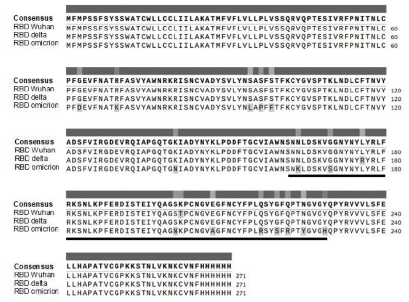
Fig. 1. Scheme of the aligned amino acid sequences of RBDs of three SARS-CoV-2 S protein mutant variants (original RBD Wuhan, RBD Delta, and the most heavily mutated RBD Omicron). Mutations of amino acid residues are highlighted in gray. The bold line represents position of the receptor-binding motif (RBM) that interacts directly with the ACE2 receptor
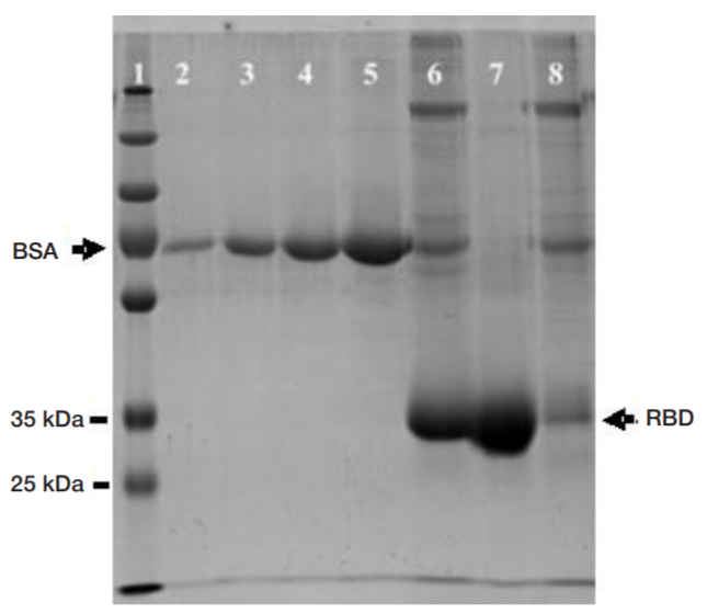
Fig. 2. SDS-polyacrylamide gel electrophoregram showing the produced (using the cloned coding sequence) and subsequently purified recombinant RBDs of three strains (6 — RBD Delta, 7 — RBD Wuhan, 8 — RBD Omicron). Left — lane of marker proteins (Thermo Scientific PageRuler Plus Prestained Protein Ladder, size 10–250 kDa). Different amounts of BSA marker protein have been applied to lanes 2–5 in order to quantify the protein produced (0.25, 0.5, 1.0, and 2.0 mg, respectively)
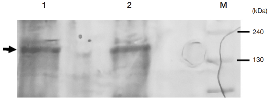
Fig. 3. SDS-polyacrylamide gel electrophoregram showing the produced SARS-CoV-2 (Wuhan) recombinant S (spike) protein. The protein was detected in the cytoplasmic fraction (1) and in the sediment fraction (2). The marker of the polypeptide molecular weight is applied on the right
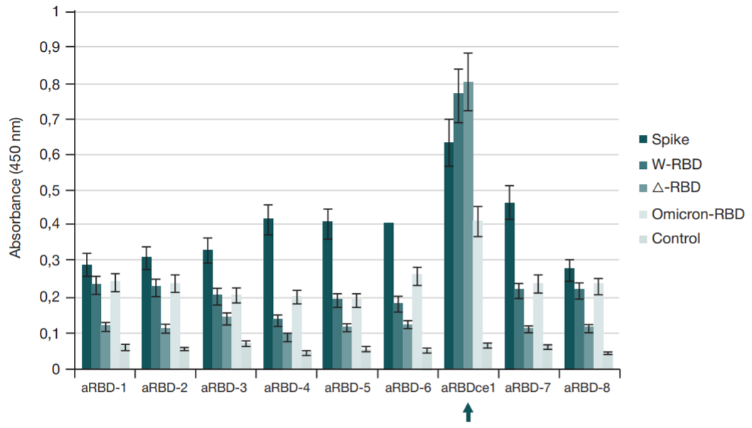
Fig. 4. Results of ELISA of nine selected nanobody variants (aRBD-1–aRBD-8 and aRBDce1) binding to immobilized recombinant proteins that correspond to (left-to-right) spike protein, RBD Wuhan (W-RBD), RBD Delta (Δ-RBD), and RBD Omicron, and to the control well with no antigen blocked with 1% BSA (as in all other wells). The absorbance values reflect the effectiveness of nanobody binding. Mean values for three independent experiments and standard deviations are provided
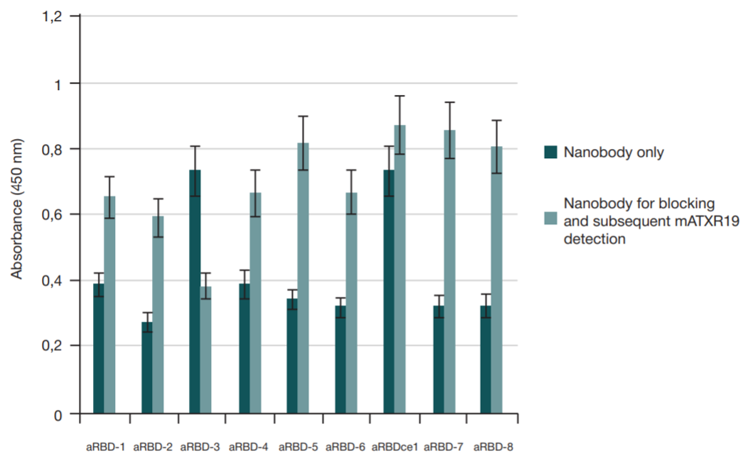
Fig. 5. Results of ELISA aimed at identification of the competition for binding to RBD Wuhan immobilized in the well between the selected nanobody variants and the XR19 neutralizing monoclonal antibody (Хеmа; Russia). The absorbance values reflect the effectiveness of nanobody binding. Mean values for three independent experiments and standard deviations are provided
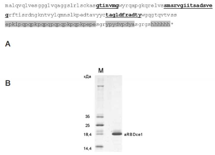
Fig. 6. аRBDсе1 nanobody. А. Amino acid sequence of the nanobody deduced from the cloned adapted coding DNA sequence. In the sequence, the CDR1, CDR2, and CDR3 hypervariable regions are highlighted (left-to-right, from N- to C-terminus) that are crucial for the coronavirus S protein RBD conserved epitope specific recognition by the аRBDсе1 nanobody. The linear linker region, HA tag, and His tag attached to the C-terminus of the nanobody are highlighted in gray. B. 14% SDS-polyacrylamide gel electrophoregram showing the produced and purified adapted аRBDсе1 nanobody
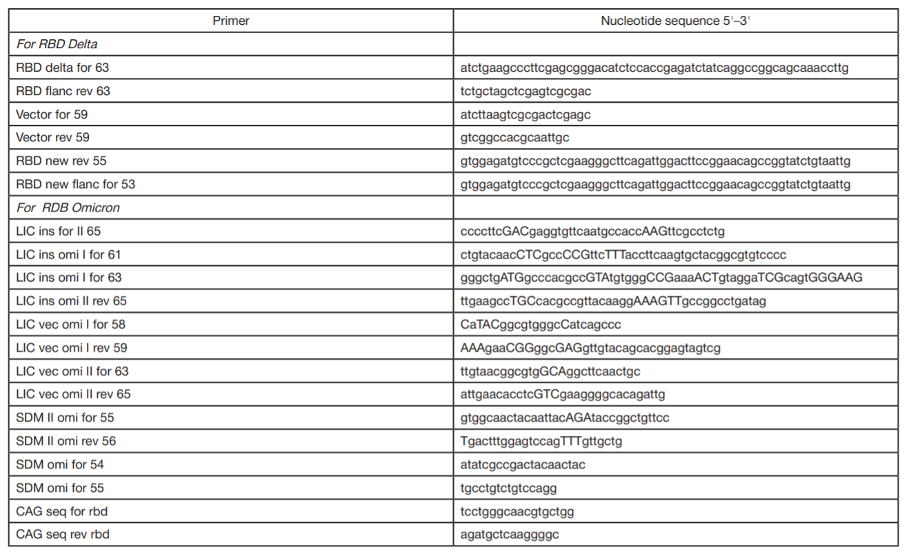
Table 1. Olygonucleotides (primers) for point mutagenesis