ORIGINAL RESEARCH

Production and biological activity of the exogenous mRNA encoding human MxA protein

Plotnikova MA1, Bobkov DE1,2, Klotchenko SA1
About authors

1 Smorodintsev Research Institute of Influenza of the Ministry of Health of the Russian Federation, St. Petersburg, Russia

2 Institute of Cytology, Russian Academy of Science, St. Petersburg, Russia

Correspondence should be addressed: Sergey A. Klotchenko
Professora Popova, 15/17, St. Petersburg, 197022, Russia; ur.liam@kitafsof

About paper

Funding: the study was supported by the Russian Science Foundation, Agreement No. 23-25-00433, project title: “Assessment of antiviral effect of the mRNA encoding human MxA protein” (manager M.A. Plotnikova), https://rscf.ru/project/23-25-00433/

Author contribution: Plotnikova MA — study design, experimental procedure, analysis of the results, statistical processing, manuscript writing; Bobkov DE — confocal microscopy examination; Klotchenko SA — production and characterization of the exogenous mRNA preparations, manuscript editing.
Received: 2024-09-30 Accepted: 2024-10-22 Published online: 2024-10-30
|
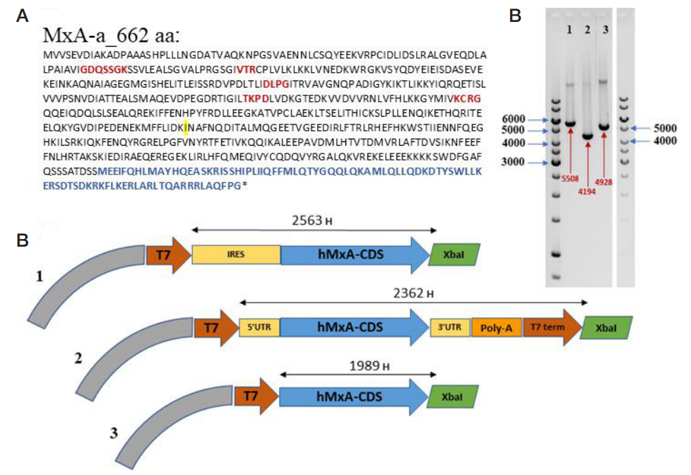
Fig. 1. Structure of the plasmid-based constructs encoding the exogenous human MxA mRNA. A. Amino acid sequence of the encoded MxA protein (obtained by sequencing of the constructs developed), the G1–G5 motifs (in the order of occurrence) are highlighted in red, the C-terminal GTPase effector domain — in blue, the V379I variation — in yellow. B. Scheme of the linearized plasmids designed for mRNA production by IVT. In the schemes, the T7 promoter region for T7 polymerase (and T7 terminator region for the construct #2) is highlighted in brick red, the auxiliary non-coding regulatory elements — in yellow, the fragment ensuring the template addition of the poly(A) tail (114 b) — in orange (in the second case), the MxA-encoding sequence — in blue, the restriction site used for linearization — in green. C. Electrophoretic separation of the linearized plasmids, the expected product lengths (bp) are highlighted in red
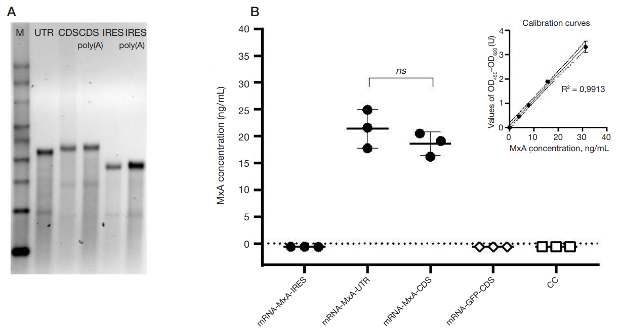
Fig. 2. Human MxA protein-encoding exogenous mRNAs and their translation in the MDCK cells. А. Electrophoretic separation of the produced exogenous mRNA products before and after polyadenylation, М — RNA molecular weight marker (#AM1750, Thermo Fisher Scientific; USA), UTR — mRNA-MxA-UTR (initially having the poly(A) tail), CDS and CDS-poly(A) — mRNA-MxA-CDS before and after polyadenylation, IRES and IRES-poly(A) — mRNA-MxA-IRES before and after polyadenylation. B. ELISA in the MDCK cells 24 h after transfection with the exogenous mRNAs
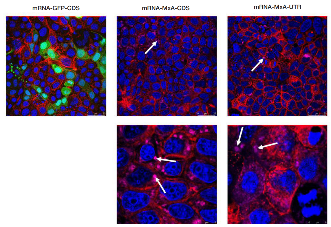
Fig. 3. Confirmation of the MxA production in the MDCK cells by confocal microscopy. Representative images of the MDCK cells (fixed specimens) were obtained 24 h after transfection with the exogenous mRNAs encoding the green fluorescent protein (mRNA-GFP-CDS) and human MxA protein (mRNA-MxA-CDS and mRNA-MxA-UTR), presented from left to right. The cell nuclei are highlighted in blue (DAPI, extinction/emission: 358 (UV)/461 nm), the actin cytoskeleton — in red (phalloidin: 540/605 nm). MxA is highlighted in magenta (620/655 nm); GFP (control cells not transfected with the MxA-encoding mRNA) is highlighted in green (488/509 nm). White arrows point to the characteristic structures defined as MxA protein
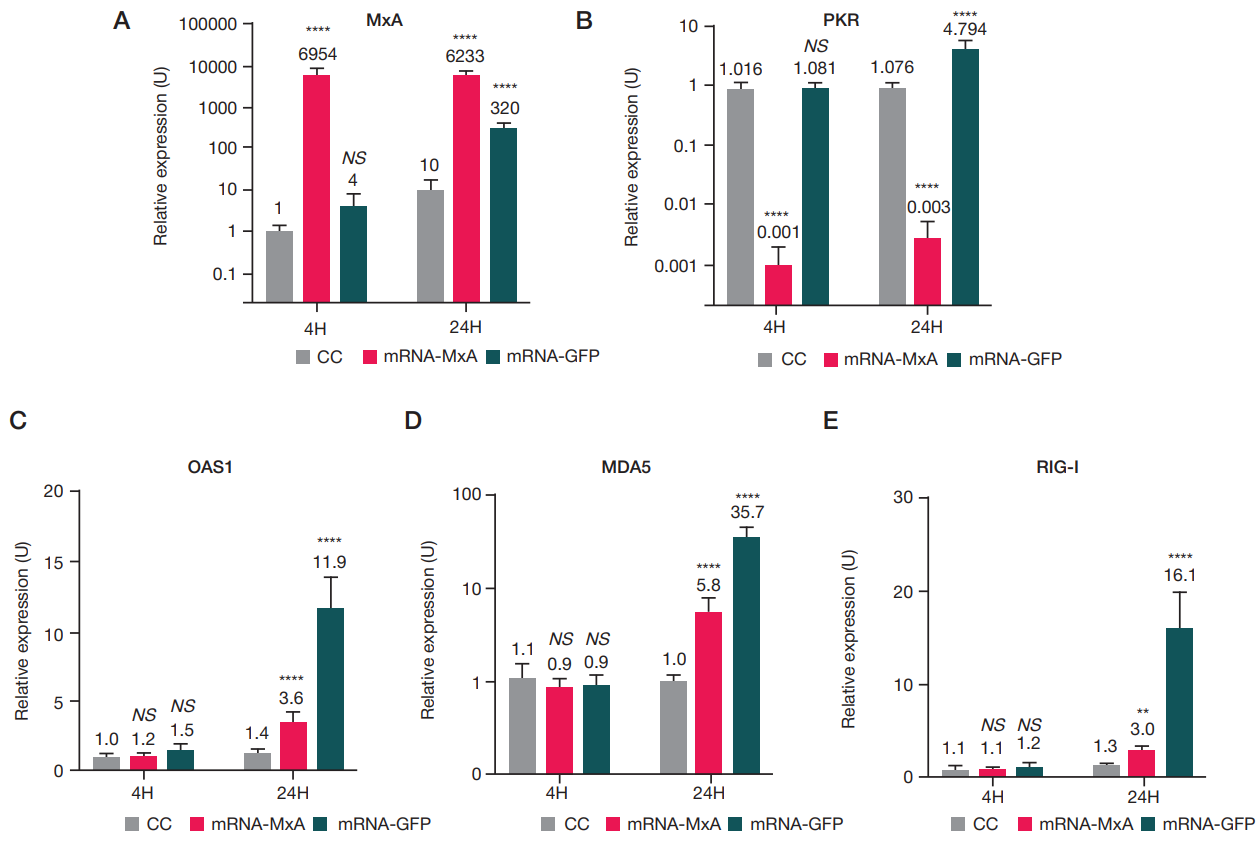
Fig. 4. Expression of the genes MxA, PKR, OAS1, as well as MDA5 and RIG-I in the A549 cells 4 h and 24 h after transfection with mRNA relative to intact cells (CC). The mean expression values (captions on top) for four biological repeats and standard error of the mean as bias are presented as bars. One-way analysis of variance (ANOVA) and the Holm–Sidak test for pairwise comparison of samples were used to calculate statistical significance of the differenced revealed. Significant differences revealed when comparing the appropriate group with the CC at the same time point are marked with asterisks: ns — Pvalue < 0.1234; * * — Pvalue < 0.0021; * * * * — Pvalue < 0.0001