ORIGINAL RESEARCH

Preliminary results of a controlled study of BCI-exoskeleton technology efficacy in patients with poststroke arm paresis

Frolov AA1,2, Mokienko OA1,3, Lyukmanov RKh1,3, Chernikova LA3, Kotov SV4, Turbina LG4, Bobrov PD1,2, Biryukova EV1,2, Kondur AA4, Ivanova GE1, Staritsyn AN1, Bushkova YuV1, Dzhalagoniya IZ2, Kurganskaya ME2,3, Pavlova OG2, Budilin SYu2, Aziatskaya GA3, Khizhnikova AE3, Chervyakov AV3, Lukyanov AL5, Nadareyshvily GG1
About authors

1 Institute of Higher Nervous Activity and Neurophysiology, RAS, Moscow, Russia

2 Pirogov Russian National Research Medical University, Moscow, Russia

3 Research Center of Neurology, Moscow, Russia

4 Vladimirsky Moscow Regional Research Clinical Institute, Moscow, Russia

5 Municipal Clinical Hospital no. 31, Moscow, Russia

Correspondence should be addressed: Olesya Mokienko
Volokolamskoye shosse, d. 80, kab. 133, Moscow, Russia, 125367; ur.xednay@dm.aysel

About paper

Funding: the study was supported by the Ministry of Education and Science of the Russian Federation (Grant Agreement no. 14.607.21.0128 dated October 27, 2015), Russian Foundation Basic Research grants no. 16-04-01506а and 16-04-00962а.
Received: 2016-03-30 Accepted: 2016-04-07 Published online: 2017-01-05
|
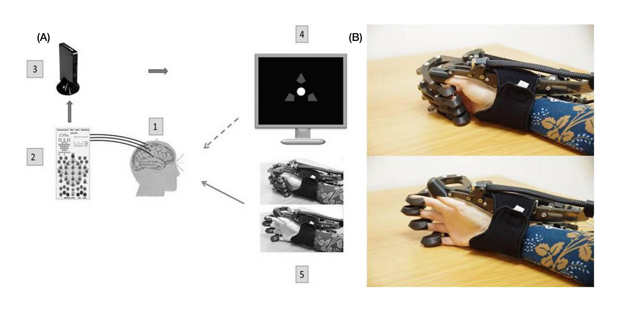
Fig. 1. BCI-exoskeleton system. (А) Schematic of the BCI used in the study: 1 — 32 Ag/AgCl EEG electrodes; 2 — NVX52 encephalograph (Medical Computer Systems, Russia); 3 — a computer (OS: Windows 7); data are transmitted in real time, EEG parameters are extracted; control command is recognized; 4 — a screen; 5 — the hand exoskeleton; dotted and solid arrows represent visual and kinesthetic feedback, respectively. (B) The hand exoskeleton (Neurobotics, Russia) with pneumatic actuator for finger extension
Fig. 2. Arm motor function dynamics in patients who completed two training courses. I and II represent the number of hospital admissions (or the training course), “Before” and “After” represent scores before and after each training course. Time elapsed after stroke with respect to the first and second hospital admissions: 21 and 30 months for Patient 1, 9 and 14 months for Patient 2, 6 and 12 months for Patient 3, respectively
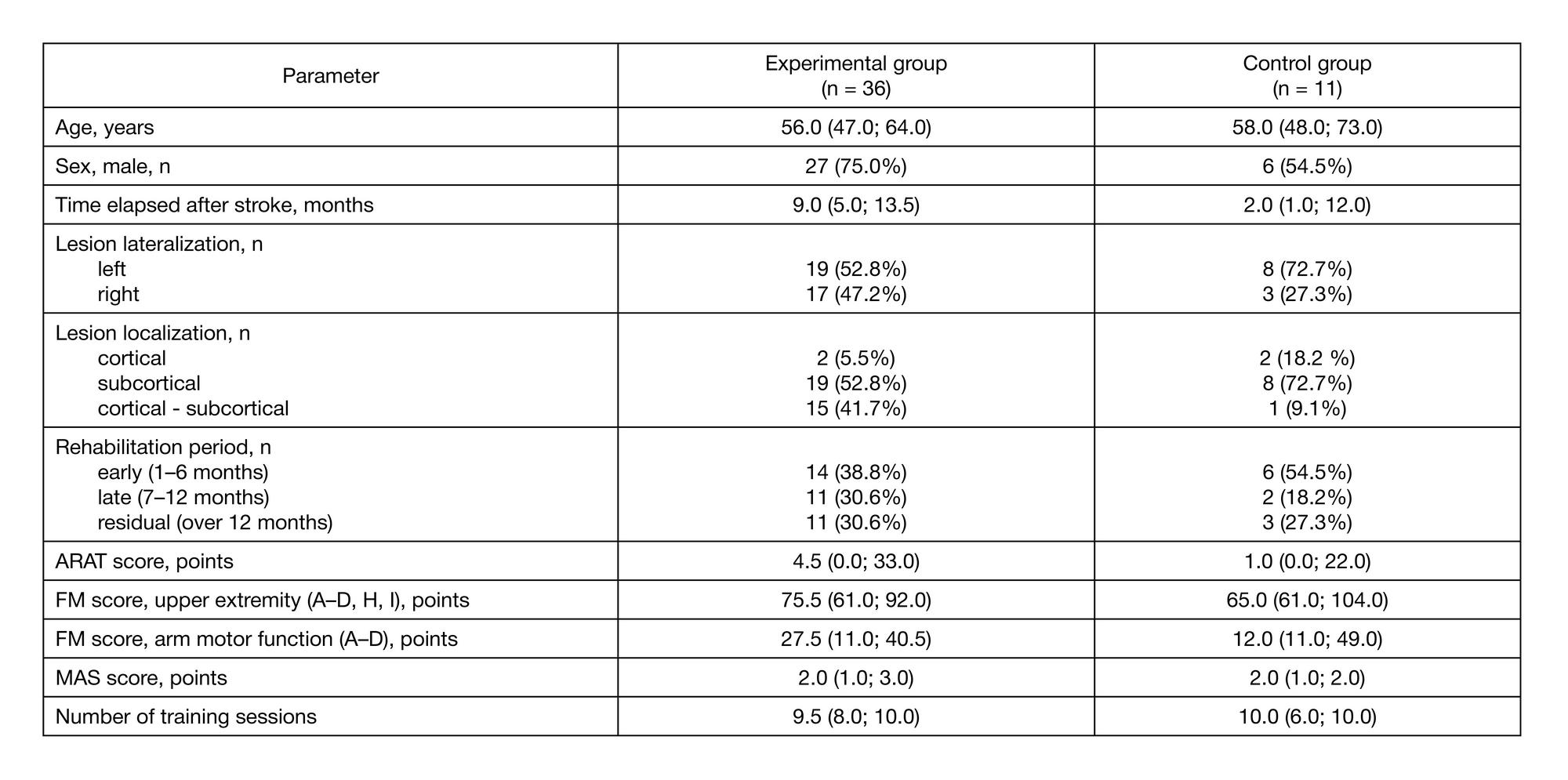
Table 1. Patients demographics and initial data (both groups)
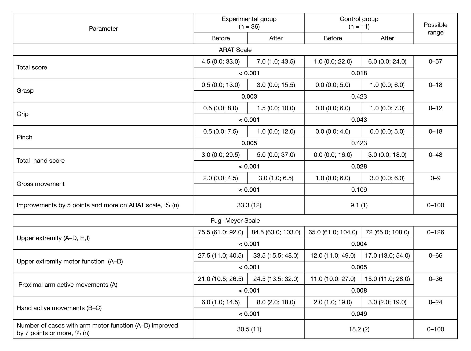
Table 2. Changes in basic ARAT and Fugl–Meyer scores in each group before and after the study
Note. Center-aligned are p values obtained from comparing the corresponding scores in each group before and after the study. Statistically significant differences are shown in bold.
Table 3. Improvement of hand motor function in the experimental group patients depending on the initial severity of paresis