ORIGINAL RESEARCH

Novel Klebsiella pneumoniae virulent bacteriophage KPPK108.1 capable of infecting the K108 serotype strains

Evseev PV1, Shneider MM1, Mikhailova YuV2, Shelenkov AA2, Yanushevich YuG3, Karlova MG4, Moiseenko AV4, Sokolova OS4, Shagin DA3
About authors

1 Shemyakin-Ovchinnikov Institute of Bioorganic Chemistry, Moscow, Russia

2 Central Research Institute of Epidemiology of Rospotrebnadzor, Moscow, Russia

3 Pirogov Russian National Research Medical University, Moscow, Russia

4 Lomonosov Moscow State University, Moscow, Russia

Correspondence should be addressed: Mikhail M. Shneider
Miklukho-Maklaya, 16/10, Moscow, 117997, Russia; ur.liam@nhs_mm

About paper

Funding: the study was funded by the Ministry of Health of the Russian Federation (EGISU R&D № 121052800048-3).

Acknowledgements: the authors wish to thank the Center for Precision Genome Editing and Genetic Technologies for Biomedicine, Pirogov Russian National Research Medical University, for advice on research methods.

Author contribution: Shagin DA — research conceptualization, study management, manuscript writing; Evseev PV, Shelenkov AA — formal analysis of sequencing data, manuscript editing; Shneider MM — methodology, study management; Mikhailova YuV — sequencing, data validation; Yanushevich YuG, Moiseenko AV, Karlova MG — methodology; Sokolova OS — electron microscopy, methodology.
Received: 2021-12-08 Accepted: 2021-12-22 Published online: 2021-12-30
|
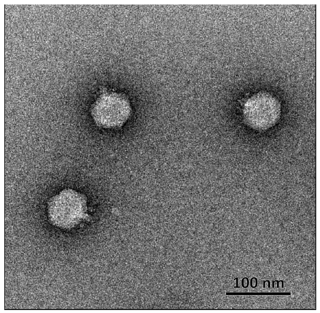
Fig. 1. Electron microscopy of Klebsiella bacteriophage KPPK108.1. Negative staining with 1% uranyl acetate, magnification ×30,000
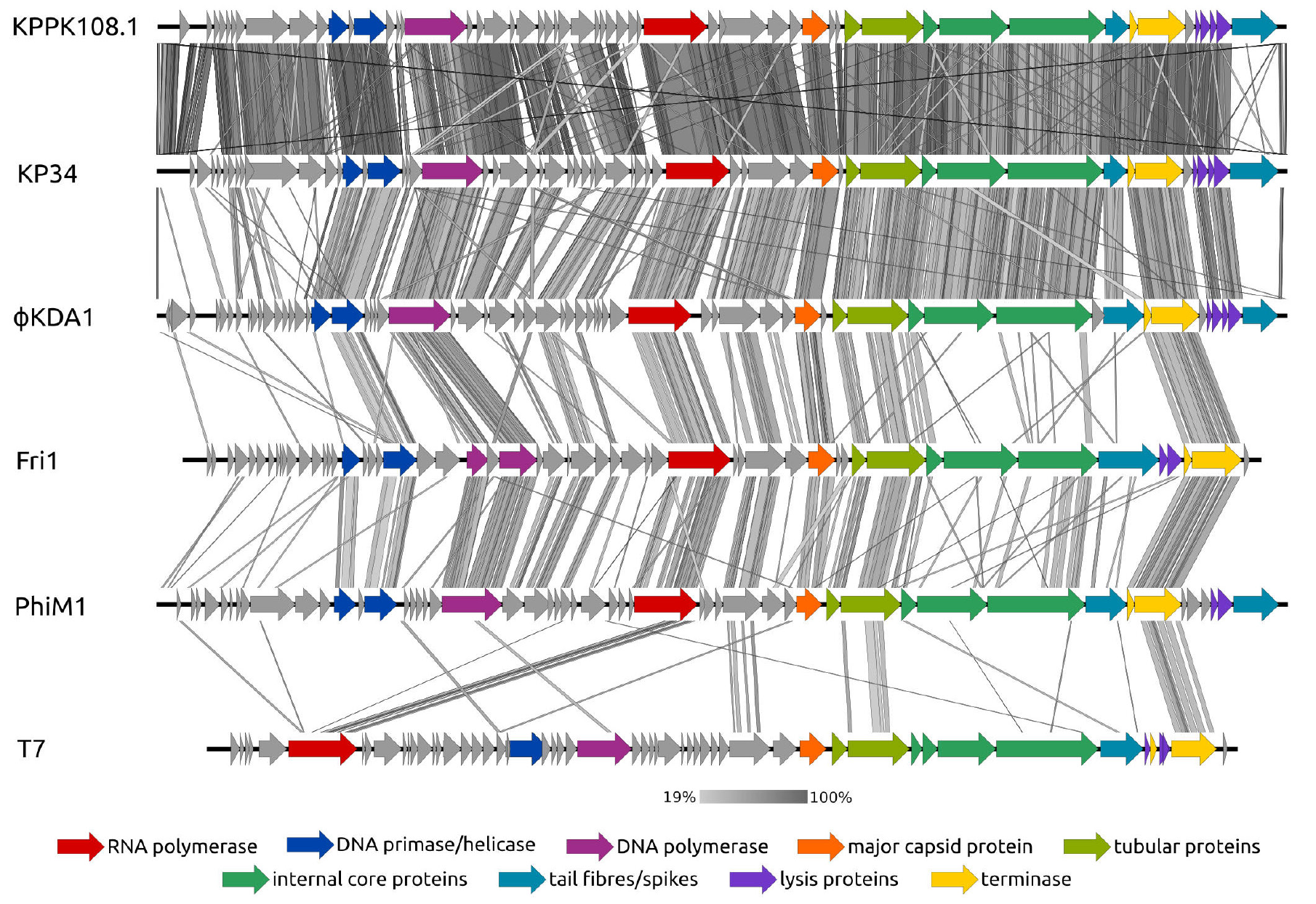
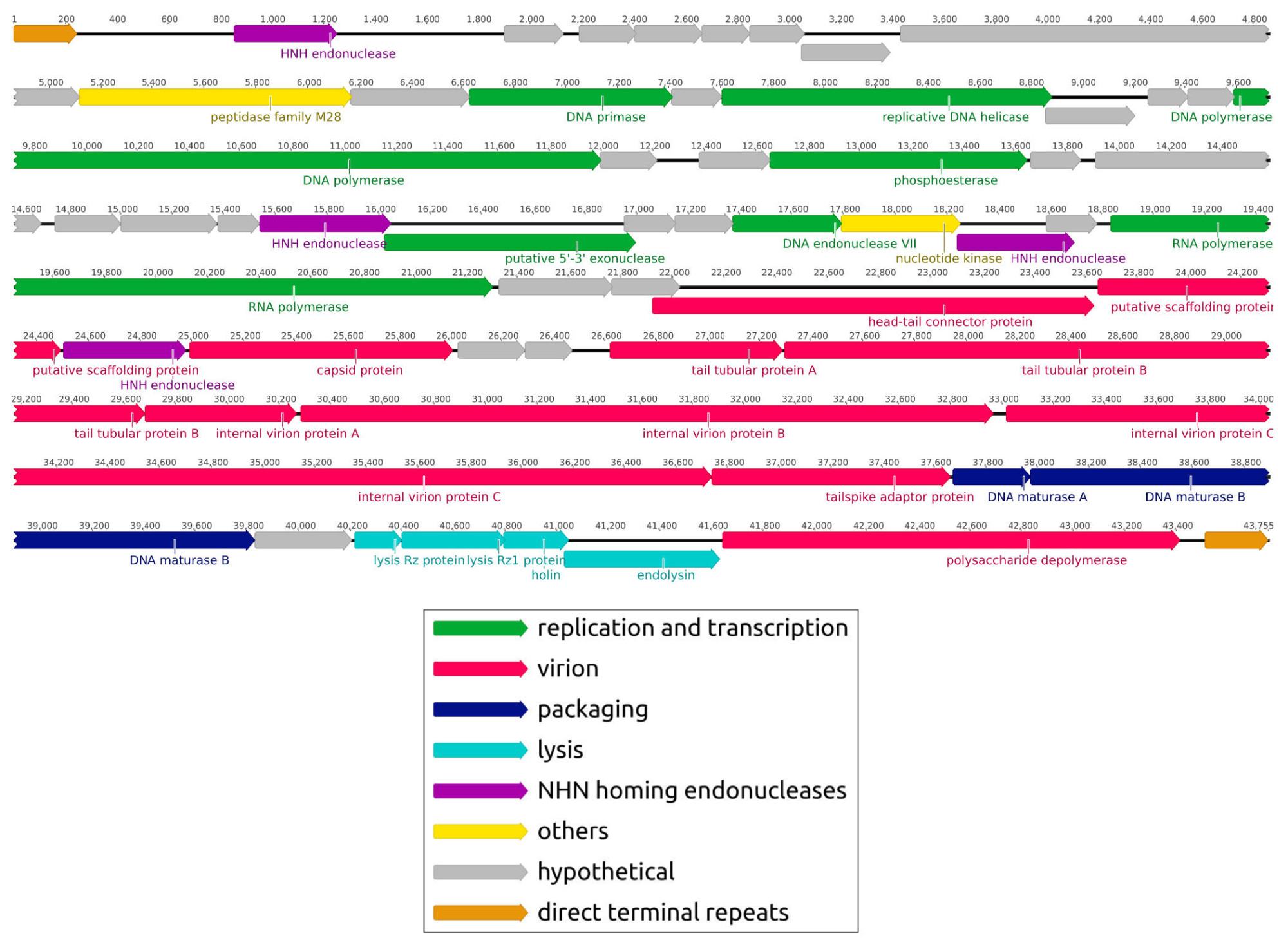
Fig. 2. Genetic map of Klebsiella bacteriophage KPPK108.1. Genes are colored in accordance with the functions of their products (see caption). Arrows indicate gene directions in accordance with their encoding functions
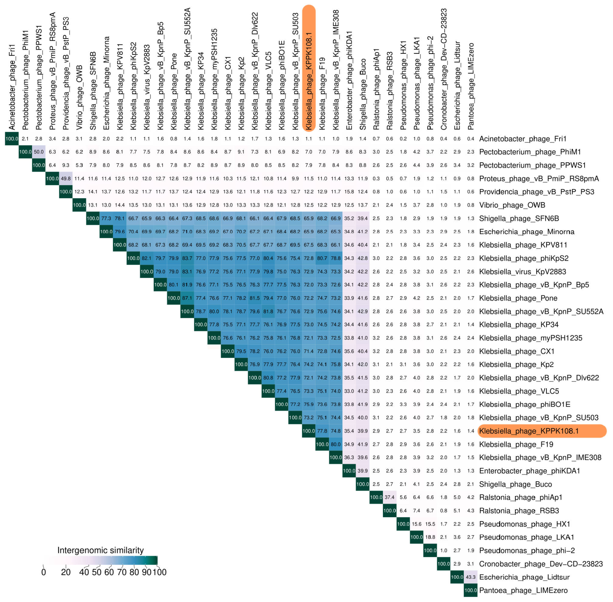
Fig. 3. Average nucleotide identity (ANI) distance matrix of the Klebsiella phage KPPK108.1 generated with the VIRIDIC web-based tool using the genomes of various Autographiviridae family members
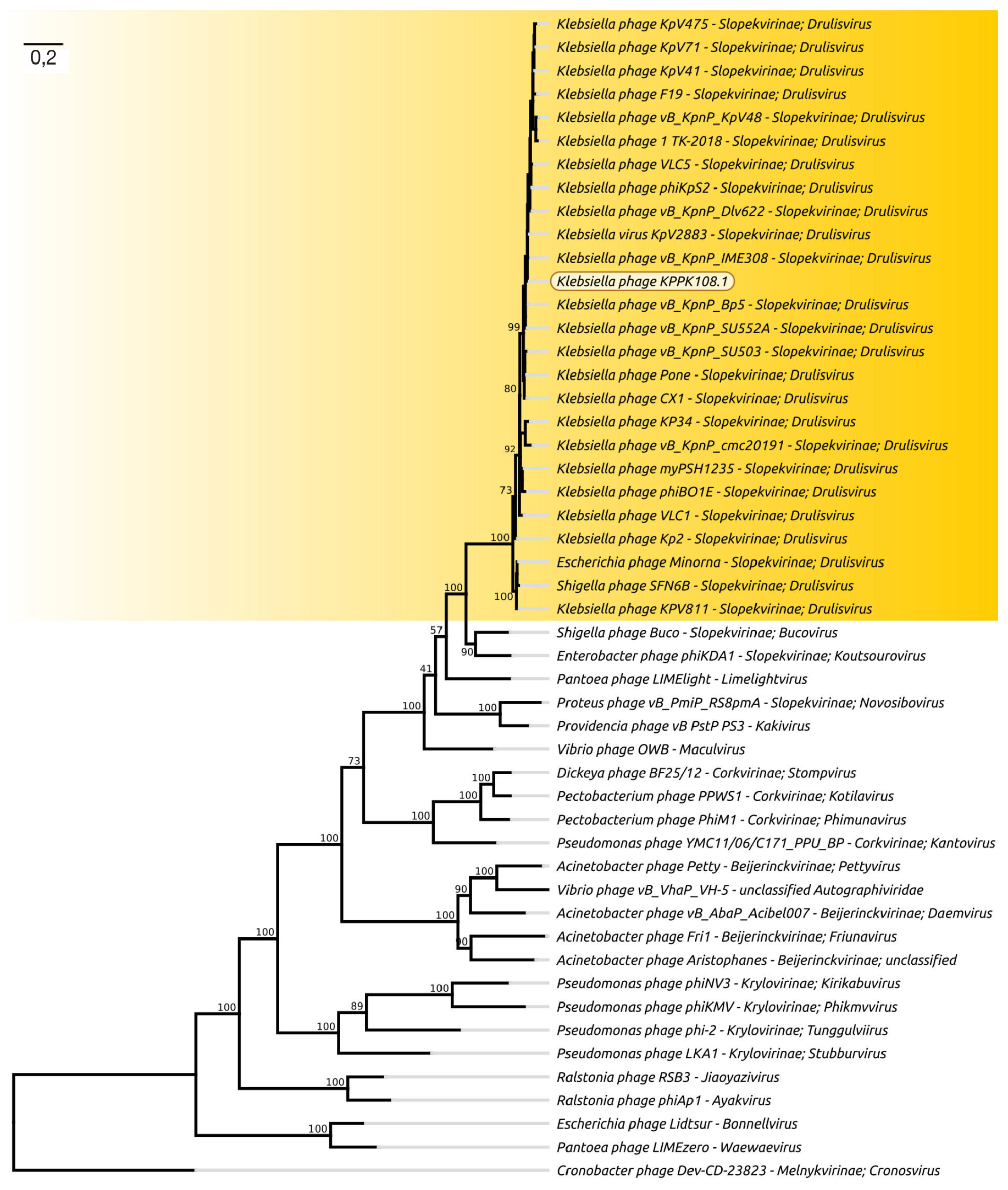
Fig. 4. Phylogenetic tree of Klebsiella phage KPPK108.1 and other Autographiviridae phages generated with the RaxML program using the concatenated amino acid sequences of the major capsid protein, terminase large subunit, DNA polymerase, and RNA polymerase
Fig. 5. Intergenomic comparison diagram created with EasyFig and TBLASTX using the genomes of Klebsiella phage KPPK108.1 and other Autographiviridae phages. The vertical lines are colored in accordance with the color scale showing the degree of similarity
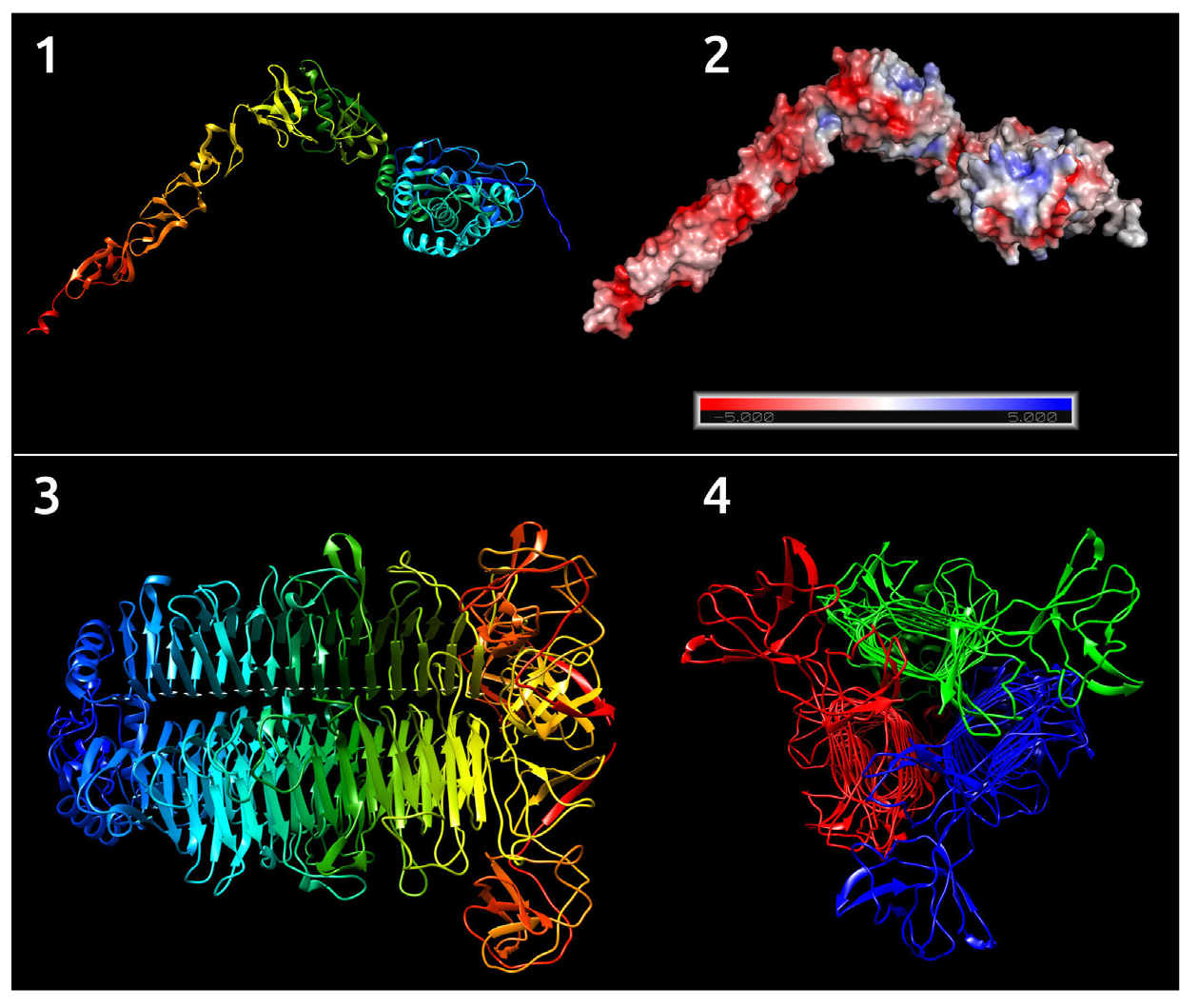
Fig. 6. Predicted tertiary structure of the KPPK108.1 phage gene 8 product painted with rainbow colors, where blue indicates N-terminal region, and red indicates C-terminal region of the protein (1). Predicted tertiary structure of the gene 8 product painted in accordance with the charge of the protein surface electrostatic field (2). Predicted tertiary structure of the KPPK108.1 phage tailspike trimer painted with rainbow colors, where blue indicates N-terminal region, and red indicates C-terminal region of the protein, longitudinal view (3). Predicted tertiary structure of the KPPK108.1 phage tailspike trimer with monomers painted with different colors, view along transverse axis (4)