ORIGINAL RESEARCH

InlB protein secreted by Listeria monocytogenes controls the pathogen interaction with macrophages

Chalenko YM1, Abdulkadieva MM2, Safarova PV1,3, Kalinin EV1, Slonova DA4, Yermolaeva SA1
About authors

1 Gamaleya National Research Center for Epidemiology and Microbiology, Moscow, Russia

2 Joint Institute for High Temperatures, Moscow, Russia

3 Pirogov Russian National Research Medical University, Moscow, Russia

4 Skolkovo Institute of Science and Technology, Moscow, Russia

Correspondence should be addressed: Yaroslava M. Chalenko
Gamaleya, 18, Moscow, 123098, Russia; ur.xednay@akazavalsoray

About paper

Funding: the study was supported by the Russian Science Foundation (project number 21-74-00105).

Author contribution: Chalenko YM — research planning, preparation and direct participation in all experiments, data interpretation and manuscript writing; Abdulkadieva MM, Safarova PV — macrophage infection assay; Kalinin EV — InlB expression analysis; Slonova DA — macrophage isolation and differentiation assay; Ermolaeva SA — research planning and manuscript writing.

Compliance with ethical standards: the research was conducted in compliance with the ethical principles of the World Medical Association Declaration of Helsinki.
Received: 2022-05-11 Accepted: 2022-06-03 Published online: 2022-06-24
|
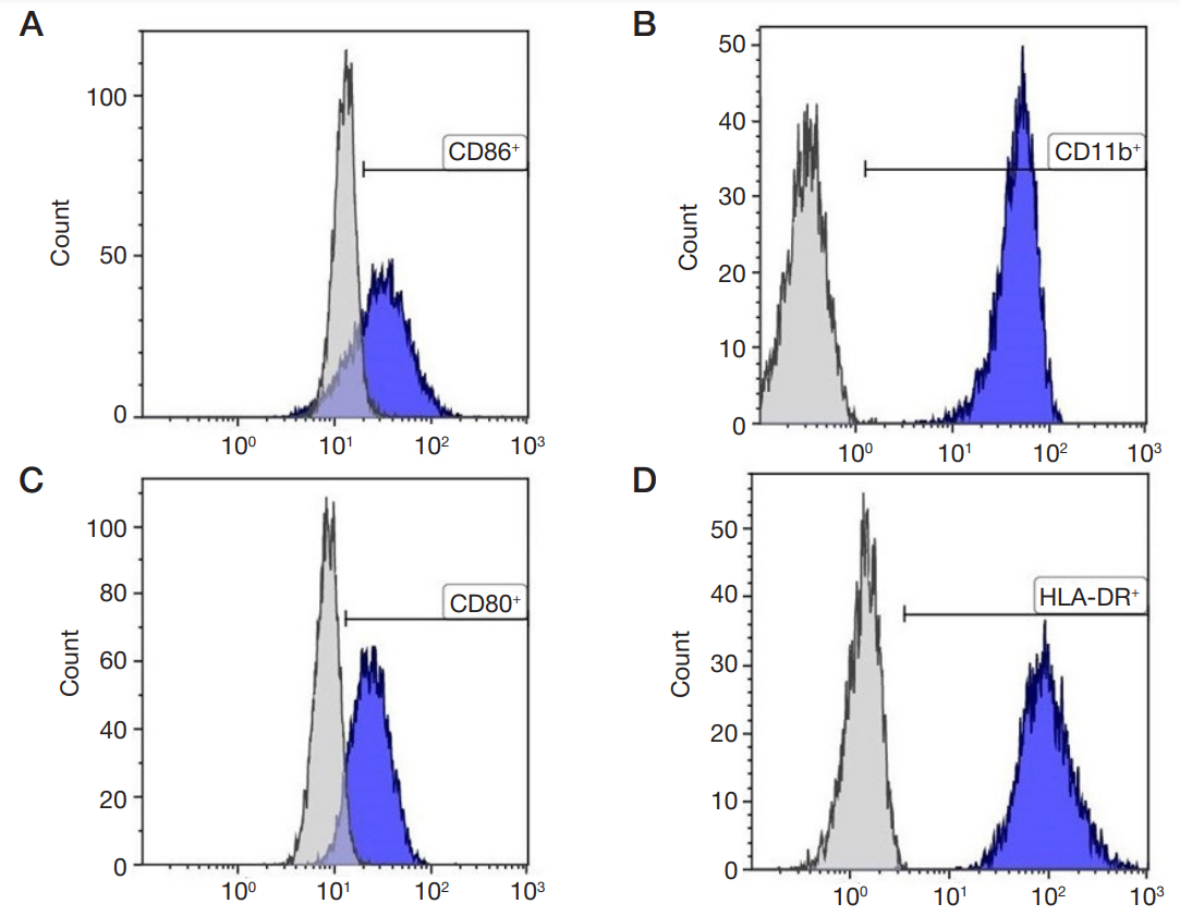
Fig. 1. Flow cytometry analysis of the macrophages obtained from peripheral blood monocytes of healthy donors. The macrophages were differentiated towards M1 phenotype; the plots show positivity for CD86 (A), CD11b (B), CD80 (C) and HLA-DR (D) markers
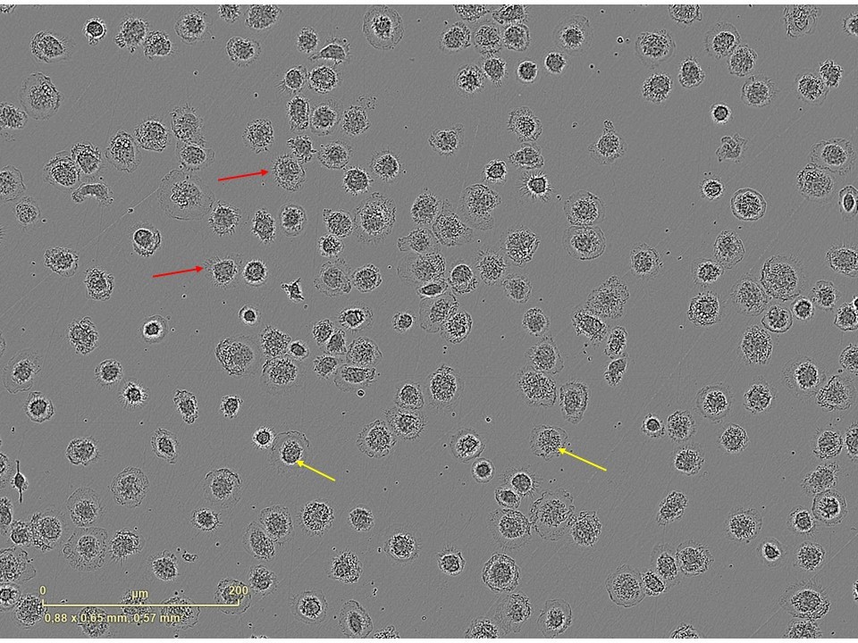
Fig. 2. Cell morphology of the M1 macrophages differentiated from peripheral blood monocytes of healthy donors. Red arrows indicate cell surface protrusions. Yellow arrows indicate large rounded nuclei. The images were acquired in an IncuCyte® S3 Live-Cell Imaging System (Sartorius; Göttingen, Germany)
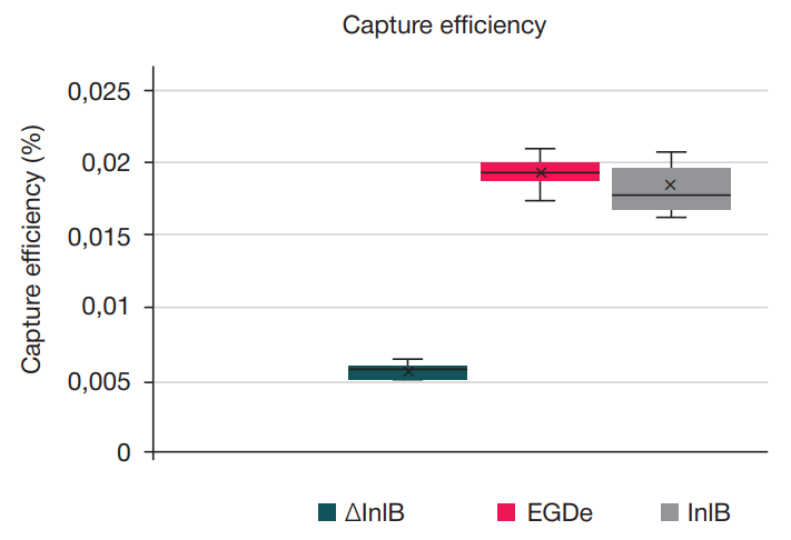
Fig. 3. Capture efficiency of L. monocytogenes ∆InlB strain, type strain EGDe and plasmid-complemented ∆InlB (InlB) strain by M1-like phenotype macrophages, ** — p < 0.01 (n = 4).
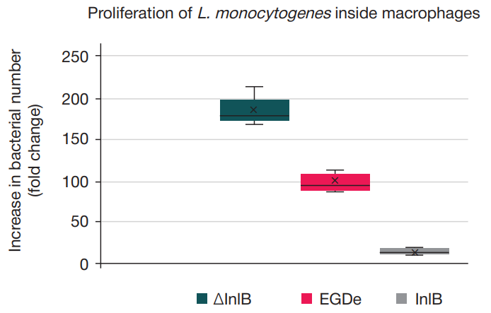
Fig. 4. Proliferation of L. monocytogenes ∆InlB strain, type strain EGDe and plasmid-complemented ∆InlB (InlB) strain inside M1-like phenotype macrophages over 24 h infection, * — p < 0,05, ** — p < 0,01 (n = 4)
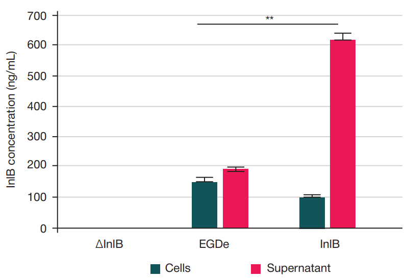
Fig. 5. Production levels of InlB protein in 18 h cultures of L. monocytogenes. The ∆InlB cultures contain no InlB protein; the type strain EGDe cultures contain 149.2 ± 13.3 ng/mL of InlB bound to the bacterial surface and 187.3 ± 9.8 ng/mL of InlB in the supernatant; the plasmid-complemented ∆InlB (InlB) cultures contain 100.7 ± 4.2 ng/mL of InlB bound to the bacterial surface and 614.6 ± 23 ng/mL of InlB in the supernatant, ** — p < 0.01 (n = 3)